Translate this page into:
Dermoscopy of tick bite
*Corresponding author: Puravoor Jayasree, Department of Dermatology, Medical Trust Hospital, Kochi, Kerala, India. dr.jayasree@medicaltrusthospital.in
-
Received: ,
Accepted: ,
How to cite this article: Jayasree P, Kaliyadan F, Ashique KT. Dermoscopy of tick bite. J Skin Sex Transm Dis 2021;3(1):103-4.
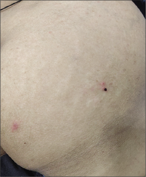
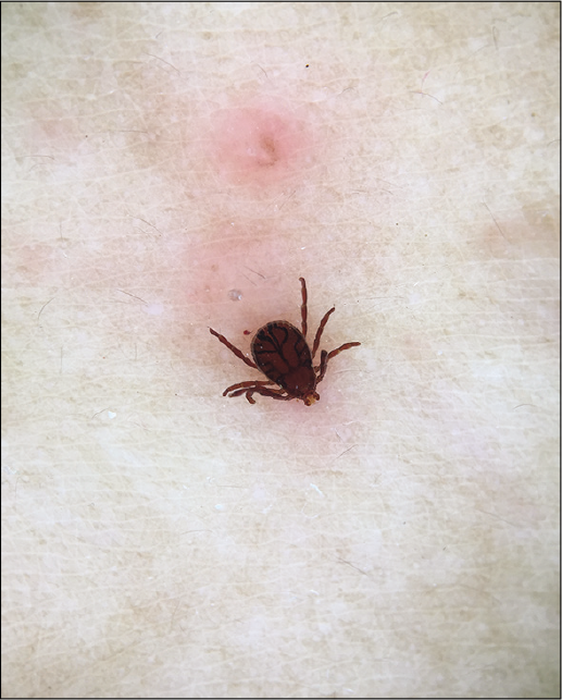
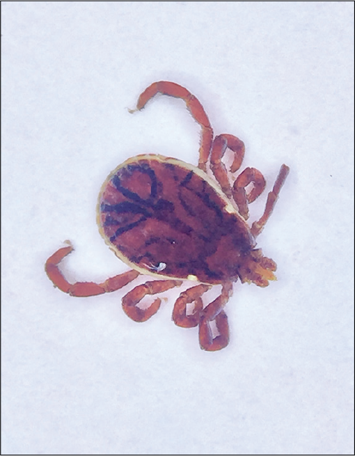
A woman in her 40s presented to our clinic with itching over her lower back and buttocks for 2 days. Examination revealed multiple discrete 1 cm sized erythematous plaques over the lower back and buttocks, with one plaque over the lateral aspect of the right buttock showing a hyperpigmented spot simulating a crust [Figure 1]. Dermoscopy revealed a live 8-legged tick still attached firmly to the erythematous area [Figure 2]. Removal of the tick was done using radiofrequency device, taking care to completely separate the tick with its mouth part intact [Figure 3].

- Discrete erythematous plaques over left buttock with one of the lesions showing hyperpigmented spot.

- Dermoscopy (Dermlite DL4N, polarized mode, ×10) showing 8-legged live tick attached to the diffuse erythematous area.

- Dermoscopy (Dermlite DL4N, polarized mode, ×10) of the 8-legged Rhipicephalus sanguineus (commonly known as brown dog tick) with its intact hypostome.
Dermoscopy of the isolated tick showed morphology consistent with Rhipicephalus sanguineus (commonly known as brown dog tick) which is known to be a vector for transmission of various Rickettsial diseases such as Indian tick typhus and ehrlichiosis. Entomodermoscopy serves as a precise diagnostic tool for tick infestations and helps in ensuring that the tick has been completely removed.
Declaration of patient consent
The authors certify that they have obtained all appropriate patient consent.
Financial support and sponsorship
Nil.
Conflicts of interest
Dr Feroze Kaliyadan and Dr Ashique KT are on the editorial board of the Journal.


