
Translate this page into:
Majocchi’s granuloma following steroid application
*Corresponding author: Geethanjali Sahadevan, Department of Dermatology, Jawaharlal Institute of Postgraduate Medical Education and Research, Karaikal, India. geethanjalisahadevan@gmail.com
-
Received: ,
Accepted: ,
How to cite this article: Mathew BP, Sahadevan G. Majocchi’s granuloma following steroid application. J Skin Sex Transm Dis. 2024;6:223-4. doi: 10.25259/JSSTD_18_2024
An 8-year-old child presented with multiple annular plaques over the trunk and limbs for a duration of two months. One of the lesions on the thigh showed follicular papules at the periphery with erosions [Figure 1]. She had been applying a steroid cream on the skin lesions for four weeks. Dermoscopy showed whitish structure-less areas, hemorrhage, polymorphic vessels, and Morse code hair [Figure 2]. In addition, she reported scrubbing the area with a loofah while bathing. A diagnosis of tinea incognito with Majocchi’s granuloma was made clinically and confirmed with a potassium hydroxide mount from the scrapings which showed septate branching hyphae of dermatophytes. She was started on oral fluconazole and topical miconazole. However, the lesion was not responding adequately, and she developed inflammatory signs such as redness and scaling. A repeat potassium hydroxide mount was done, which showed fungal filaments. She was not willing to undergo a biopsy. The child was started on oral terbinafine at a dose of 125 mg/day, and within a week, the lesion started to regress and completely subsided in three weeks.
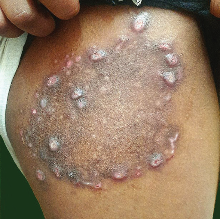
- Clinical image showing annular plaque with peripheral follicular papules.
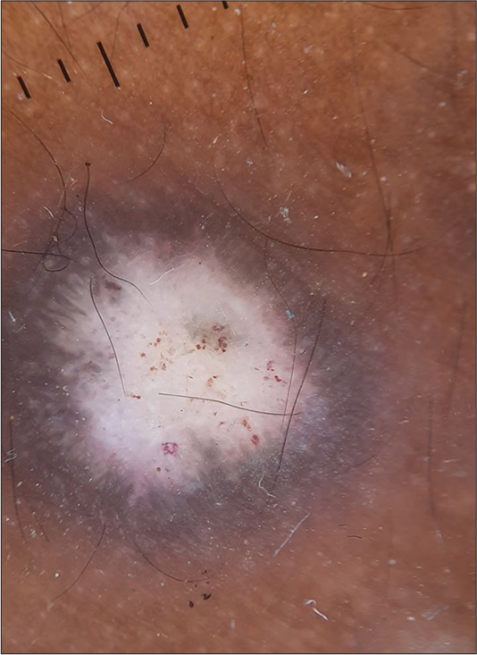
- Dermoscopy image showing white structure-less areas, hemorrhage, polymorphous vessels, and Morse code hair (DermLite DL4 ×10).
Ethical approval
The Institutional Review Board approval is not required.
Declaration of patient consent
The authors certify that they have obtained all appropriate patient consent.
Conflicts of interest
There are no conflicts of interest.
Use of artificial intelligence (AI)-assisted technology for manuscript preparation
The authors confirm that there was no use of artificial intelligence (AI)-assisted technology for assisting in the writing or editing of the manuscript and no images were manipulated using AI.
Financial support and sponsorship
Nil.


