Translate this page into:
Dermoscopy of lipoid proteinosis – Topographic diverse features
*Corresponding author: Arun Somasundaram, Department of Dermatology and STD, Jawaharlal Institute of Postgraduate Medical Education and Research, Puducherry, India. arunsomasundaram25@gmail.com
-
Received: ,
Accepted: ,
How to cite this article: Somasundaram A. Dermoscopy of lipoid proteinosis – Topographic diverse features. J Skin Sex Transm Dis. 2024;6:210-2. doi: 10.25259/JSSTD_69_2023
Dear Editor,
Lipoid proteinosis (LP), known as Urbach–Wiethe disease or hyalinosis cutis et mucosae, is a rare disease, with approximately 400 cases reported in the literature, equally affecting males and females. It is an autosomal recessively inherited genodermatosis due to mutations in the extracellular matrix protein gene (ECM1) located on chromosome 1q21.2.[1,2] Deposition of hyaline material in the mucosae, skin, and brain tissue results in persistent hoarseness from infancy. It is accompanied by skin changes, such as fragility, as evident by blistering, infiltrated papules, and nodules. Affected individuals may also present with neurologic and psychiatric symptoms. It is crucial to be aware of these myriad manifestations for managing these patients. Dermoscopy plays a vital role in assisting the diagnosis. We report a case of LP with site-specific dermoscopic observations.
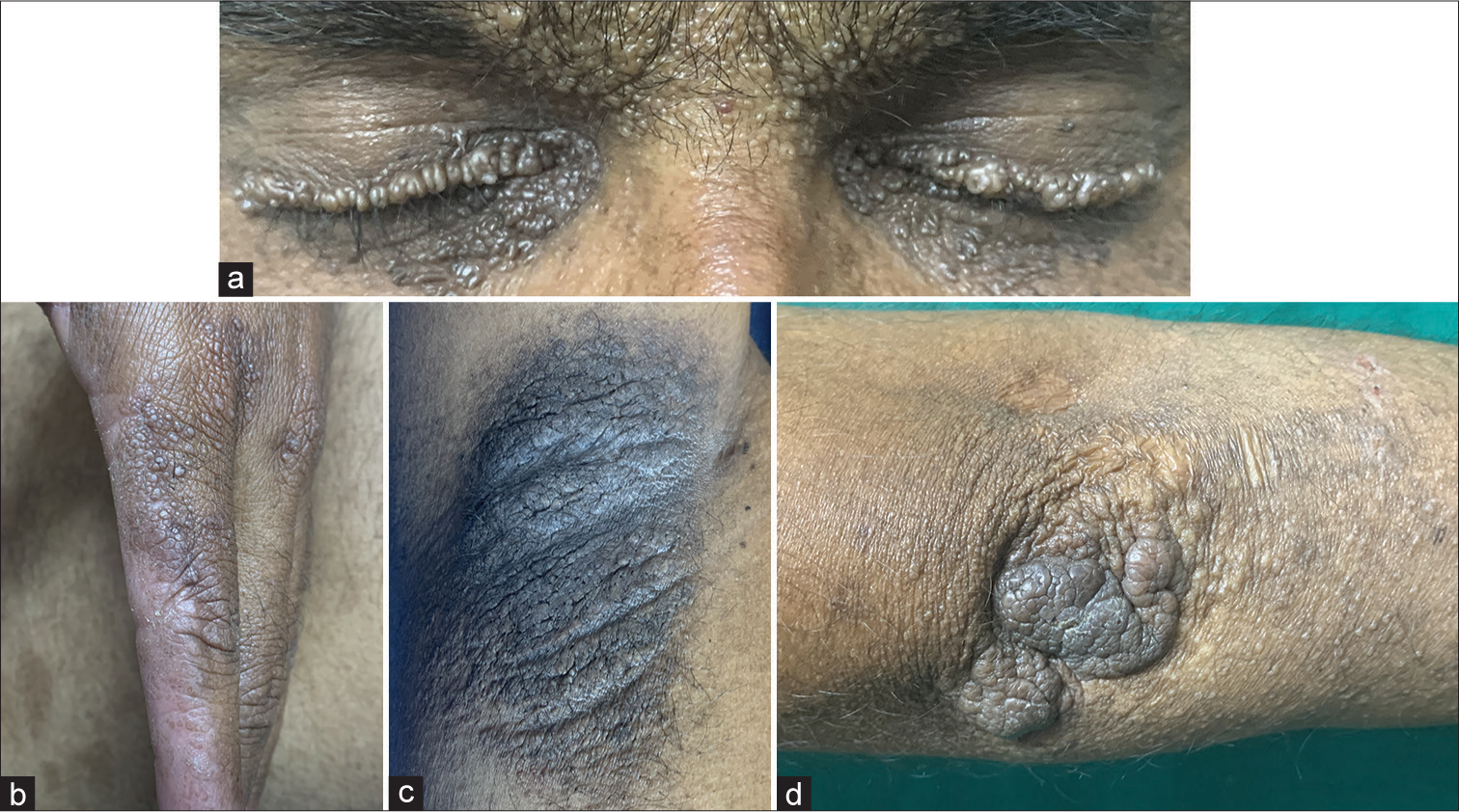
A 35-year-old male patient, the second born of third-degree consanguinity, noticed a history of itchy blisters over his face, trunk, and extremities that healed with pock-like scars. He gave a history of similar complaints in his female sibling. There was hoarseness of voice noticed from birth in both of them. Cutaneous examination revealed beaded papules along eyelid margins suggestive of moniliform blepharosis [Figure 1a] significant varioliform scars over the face, trunk, and extremities, difficulty in protruding the tongue outside and grouped skin-colored papules over the sides of his fingers [Figure 1b]. He also had verrucous plaques over his axilla [Figure 1c] and elbows [Figure 1d]. Oral cavity and genitalia were normal. Dermoscopy revealed different findings at various sites. DermLite DL4 (×10, polarized mode) showed clustered whitish-yellow clods in a linear configuration over the beaded papules with distichiasis evident along the eyelid margin [Figure 2a] and pearly beaded globules over the side of fingers [Figure 2b]. Dermoscopy also revealed sulci-gyri appearance over verrucous plaque over the axilla [Figure 2c] and grayish-white rounded structures in clumps, giving a pulpy appearance over the elbow [Figure 2d]. Our patient did not have any other systemic involvement. Acitretin was started after a pre-retinoid workup, and the patient was counseled regarding the disease, the need for follow-up, and the prognosis. Skin biopsy was not done on our patient as our patient did not consent to the same, and we managed the patient with our diagnosis clinically as he had classical features with a family history. Hence, a dermoscopy histopathology correlation was not done in our case. There are anecdotal reports on dermoscopy of LP. Our case highlights the site-specific dermoscopic features that were interesting to report.[1,2] In our patient, sulci and gyri predominated over the skin lesions of the elbow and axilla, while whitish-yellow clods and pearly beaded globules correspond to the amyloid deposits. Dermoscopy features described in the literature include clustered pale structureless globules in a linear configuration along the upper and lower palpebral margin with misaligned eyelashes and pearly beaded globules corresponding to amyloid deposition in the dermis. The sulci with gyri appearance in the verrucous plaques leading to cerebriform appearance correspond to hyperkeratosis with papillomatosis and elongated rete ridges on histopathology.[1,2] Dermoscopy can sometimes help diagnose the beaded papules along the lid margins when the findings are subtle in some cases.[2] The early cutaneous features sometimes may be elusive, and in such a scenario, dermoscopy may aid as a shred of supportive evidence to the diagnosis.

- (a): Beaded papules over eyelids suggestive of moniliform blepharosis; (b): Beaded skin-colored papules over the sides of fingers; (c): Verrucous plaque over axilla; (d): Verrucous plaque over the elbow.

- Dermoscopy – DermLite (×10, polarized mode) (a): Eyelids – Multiple whitish-yellow clods in a linear configuration over beaded papules with distichiasis; (b): Pearly beaded globules over the sides of fingers; (c): Axilla-sulci and gyri give a cerebriform appearance; (d): Elbows – Grayish-white rounded structures in clumps giving a pulpy appearance. The blue arrows (a-d): describes the dermatologic findings.
Ethical approval
Institutional Review Board approval is not required.
Declaration of patient consent
The authors certify that they have obtained all appropriate patient consent.
Conflicts of interest
There are no conflicts of interest.
Use of artificial intelligence (AI)-assisted technology for manuscript preparation
The authors confirm that there was no use of artificial intelligence (AI)-assisted technology for assisting in the writing or editing of the manuscript and no images were manipulated using AI.
Financial support and sponsorship
Nil.
References
- Novel features in dermoscopy of lipoid proteinosis. Indian J Dermatol Venereol Leprol. 2023;89:116-8.
- [CrossRef] [PubMed] [Google Scholar]
- Lipoid Proteinosis with esotropia: Report of a rare case and dermoscopic findings. Indian J Dermatol. 2020;65:53-6.
- [CrossRef] [PubMed] [Google Scholar]


