Translate this page into:
Looking at the “Inverse” in dermatology
*Corresponding author: Pooja Bains, Associate Professor, M.D. (Skin & VD), Department of Dermatology, Sri Guru Ram Das Institute of Medical Sciences and Research (SGRDIMSR), Amritsar, Punjab, India. pjdhawan76@gmail.com
-
Received: ,
Accepted: ,
How to cite this article: Bains P. Looking at the “Inverse” in dermatology. J Skin Sex Transm Dis 2024:6:66-70. doi: 10.25259/JSSTD_46_2023.
Abstract
The predilection of skin disorders for characteristic sites and awareness of these sites is very important for clinical diagnosis. However, certain subtypes of common dermatological conditions can involve sites opposite to that of the classical presentation. These subtypes are named using the prefix “inverse” or the suffix “inversus/inversa” in the literature. These variants may pose a diagnostic challenge to residents and knowledge of the various “inverse” dermatological conditions and phenomena in the literature is of great significance.
Keywords
Inverse
Inversus
Inversa
Inversum
INTRODUCTION
There are certain subtypes of common dermatological conditions where the distribution is opposite to the classical distribution. These subtypes are named using the prefix “inverse” or the suffix “inversus/inversa” in the literature. The Latin words “inversus” (masculine) and “inversa” (feminine) meaning “upside down” are used for these dermatological conditions. An electronic and manual search of standard dermatology textbooks and databases including PubMed/Medline and Google Scholar was done to include all the “inverse” variants of skin diseases. These uncommon to rare inverse variants add to the diverse presentation in dermatological diseases.
INVERSE PSORIASIS
This subtype (also referred to as flexural or intertriginous psoriasis) commonly involves the groins, axillae, inframammary creases, perianal, and retroauricular regions. In rare cases, it can involve the antecubital and popliteal fossae and the interdigital spaces.[1,2] The plaques of Inverse Psoriasis are thin and well-defined, with reduced or absent scaling and a glazed surface as shown in Figures 1 and 2.[3] There can be a sudden appearance of Inverse Psoriasis in HIV-infected adults.[4]
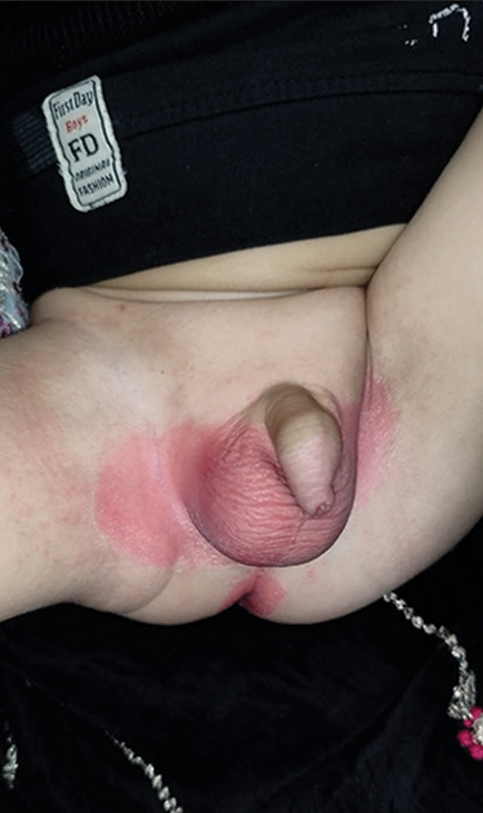
- Napkin psoriasis in an infant.
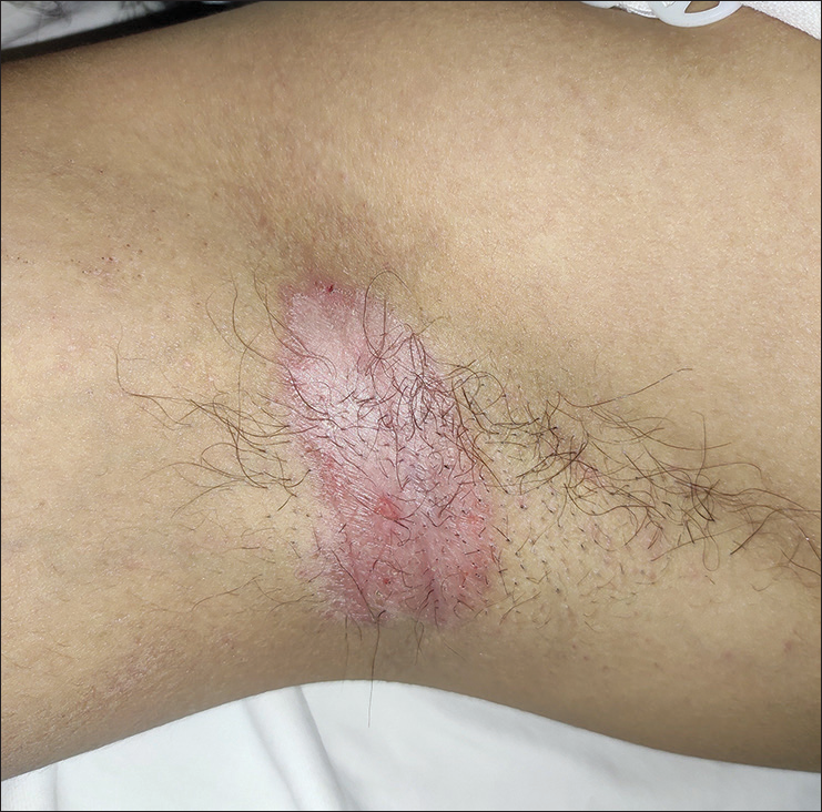
- Inverse psoriasis in an adult involving axilla.
INVERSE PITYRIASIS ROSEA/PITYRIASIS ROSEA INVERSUS
When the lesions of pityriasis rosea spare the trunk and affect the axillary and inguinal folds, face, neck, and extremities, the variant is called inverse pityriasis rosea [Figure 3].[5] It is prevalent in the pediatric age group and in dark-skinned individuals.[6]
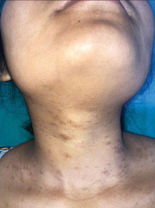
- Inverse pityriasis rosea in a young female involving the neck.
INVERSE LICHEN PLANUS (LP)
This is an uncommon variant of LP in which the characteristic violaceous papules are seen in the axillary and inguinal folds and the inframammary areas. It can also involve other flexural areas such as the popliteal and antecubital fossae.[7] The lesions of inverse LP present as non-scaly erythematous lesions with ill-defined borders. In addition, erosions, hyperpigmentation, and hyperkeratotic papules can occur.[8]
LICHEN PLANUS PIGMENTOSUS (LPP) INVERSUS
Pock et al. described this rare subtype of LP in 2001.[9] LPP inversus is characterized by mildly itchy hyperchromic macules in the flexural areas, mainly the axillary folds (90%) and groins, and rarely, the antecubital and popliteal areas.[10] There is an absence of involvement in sun-exposed areas in contrast to classical LPP. The closest differential is ashy dermatosis which has a predilection for the trunk and limbs, and not the flexural areas.[11]
INVERSE GOTTRON PAPULES
Gottron’s papules which are a characteristic sign of dermatomyositis are inflammatory, flat-topped, lichenoid papules on the extensor aspect of hands, particularly over the interphalangeal and metacarpophalangeal joints of the fingers.[12] When these papules are present on the palmar surfaces of the finger creases as hyperkeratotic papules, they are referred to as inverse Gottron’s papules.[13] Inverse Gottron papules have been associated with severe Interstitial lung disease in adults and anti-MDA5 antibodies.[14]
ACNE INVERSA
The term “acne inversa” was proposed in 1989 by the Plewig and Steger for hidradenitis suppurativa (HS).[15] The term “acne inversa” indicates the involvement of pilosebaceous unit in the disease.[16] The histopathology and pathogenesis are similar to classical acne while the involvement of intertriginous sites is in contrast to acne. Some dermatologists are of the opinion that the term “acne inversa” should be preferred over “hidradenitis suppurativa.”[17] In contrast, some are of the view that HS, though a misnomer, identifies a distinct entity that has discrete genetic basis, etiological factors, and disease associations, and therefore, the name should not be replaced.[18]
PTERYGIUM INVERSUM UNGUIS (PIU)
This is a rare condition which affects the distal part of the nail bed. The hyponychium is attached to undersurface of the nail plate. Therefore, with the growth of the nail plate, the hyponychium extends forward resulting in expungement of the distal groove.[19] In classical pterygium unguis, the proximal nail fold fuses with the nail matrix and nail bed. PIU is otherwise called ventral pterygium. It can be congenital or acquired.[20] The acquired forms may be idiopathic or consequential to systemic conditions such as connective tissue diseases, leprosy, neurofibromatosis, or paresis.[21] Figure 4 shows pterygium inversum unguis in a patient with systemic sclerosis.
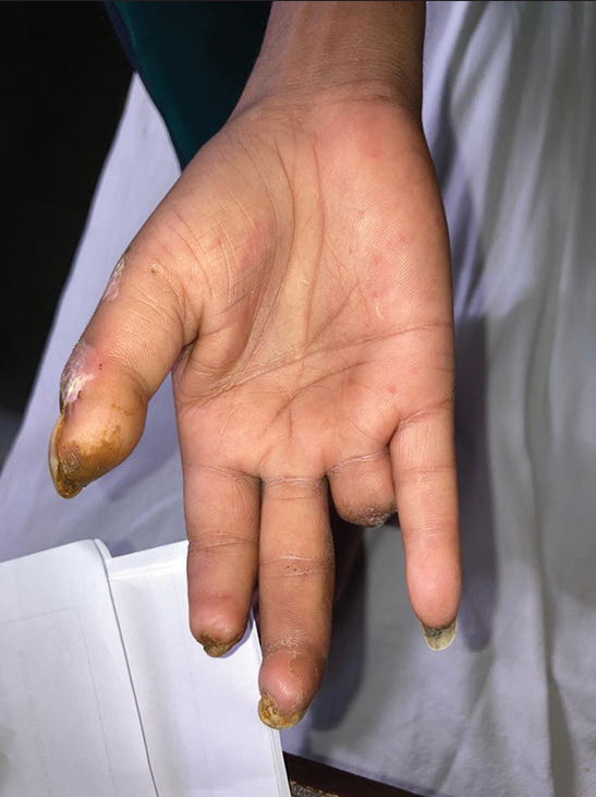
- Pterygium inversum unguis in a patient of systemic sclerosis.
OPHIASIS INVERSUS
Muñoz and Camacho published the first description of this variant of alopecia areata in 1996. It is also known as sisaipho (ophiasis spelled backward).[22] It presents with non-scarring alopecia sparing the temporal and occipital areas [Figure 5]. Various studies have shown its increased association with atopy, vitiligo, thyroid disease, and trachyonychia.[23] The prognosis of this condition is better that of ophiasis.[23]
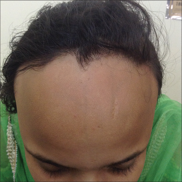
- Ophiasis inversus.
INVERSE TYPE OF JUNCTIONAL EPIDERMOLYSIS BULLOSA (JEB INVERSA)
JEB inversa is a rare variant with symmetrical involvement of the body folds, that is, the axillary, inguinal, and perineal areas. The erosions heal with atrophic white scars. It has a non-lethal course with generalized blistering in the neonatal period and only flexural involvement in adult life. The proposed hypothesis is that some mutant proteins might be more unstable in the warmer regions of temperatures of the body folds.[24]
INVERSE TYPE OF RECESSIVE DYSTROPHIC EPIDERMOLYSIS BULLOSA (RDEB-I)
This is a rare variant of RDEB which presents in infancy with blistering, erosions, and atrophy over the axillae, thighs, groins, and neck. In adulthood, there is predominant involvement of the perineal area and the axillary folds.[25] Hands, feet, elbows, and knees are not involved.[26] This type of EB was first described in 1971 by Gedde Dahl.[27] It frequently involves the oral and esophageal mucosae, cornea, vulva, external ear canal, and nails. There is an increased risk of dental caries.[28] A study observed that Collagen7 from RDEB-I mutations demonstrated instability at high temperatures.[29] RDEB inversa carries a comparatively better prognosis.[27]
INVERSE KLIPPEL-TRENAUNAY SYNDROME
The classical features of Klippel-Trenaunay syndrome include capillary malformation of skin (port wine stain) with ipsilateral soft tissue and bone hypertrophy, and varicose veins. In the inverse presentation, the affected limb has deficient growth and is smaller in size. Although it has been postulated that the presence of a minus allele might be responsible for this deficient growth, the cause is still unknown.[30]
INVERSE PAPULAR ACROKERATOSIS (ACROKERATOELASTOIDES OF OSWALDO COSTA)
It is a rare genodermatosis presenting with skin-colored, horny papules on the lateral and dorsal aspects of palms and soles. It was described by Oswaldo Costa in 1952 and is seen mainly in young dark skinned women as asymptomatic keratotic papules on the lateral margins of hands and feet.[31]
There are a few more case reports of “inverse” conditions as summarized in Table 1.[32-39]
Table 2 enumerates Inverse phenomena and appearances in dermatology.[40-45]
| Inverse Koebner phenomenon/Renbok phenomenon | First reported by Happle et al. in 1991 to describe normal hair growth seen in a psoriatic plaque in a patient with alopacia areata of the scalp.[40] The cellular alteration and change in the microenvironment by the first injury prevent the development of subsequent disease at the same site.[41] |
| Inverse halo phenomenon | Loss of pigmentation starts from the core of a melanocytic nevus rather than at the periphery.[42] |
| Inverse shouldering effect | The characteristic appearance seen in lipedema is present in females. Other names for this appearance are armchair appearance, bracelet sign, and cliff sign.[43] There is a symmetric increase in subcutaneous fat in bilateral lower limbs which stops abruptly at the level of malleoli and spares the feet.[44] |
| Inverse pigmentary network pattern | The reverse of the pigment network pattern on dermoscopy comprises white/lighter lines forming the grid of the network with comparatively darker areas filling the gaps. Other terms used for this pattern are negative pigment network and reversed pigment network. It is seen in melanoma, dermatofibroma, Spitz naevus, and evolving lesions of vitiligo.[45] |
CONCLUSION
The involvement of sites opposite to the classical disease might pose a diagnostic challenge to a dermatology resident. Hence, knowledge of these unusual subtypes and their recognition is of utmost importance.
Ethical approval
The Institutional Review Board approval is not required.
Declaration of patient consent
The authors certify that they have obtained all appropriate patient consent.
Conflicts of interest
There are no conflicts of interest.
Use of artificial intelligence (AI)-assisted technology for manuscript preparation
The author(s) confirms that there was no use of artificial intelligence (AI)-assisted technology for assisting in the writing or editing of the manuscript and no images were manipulated using AI.
Financial support and sponsorship
Nil.
References
- Psoriasis and related disorders In: Griffiths C, Barker J, Bleiker T, eds. Rook’s textbook of dermatology (9th ed). Oxford: Blackwell Science; 2016. p. :35.1-48.
- [Google Scholar]
- Psoriasis inversa: A separate identity or a variant of psoriasis vulgaris? Clin Dermatol. 2015;33:456-61.
- [CrossRef] [PubMed] [Google Scholar]
- Inverse psoriasis: From diagnosis to current treatment options. Clin Cosmet Investig Dermatol. 2019;12:953-9.
- [CrossRef] [PubMed] [Google Scholar]
- HIV-associated psoriasis: Pathogenesis, clinical features, and management. Lancet Infect Dis. 2010;10:470-8.
- [CrossRef] [PubMed] [Google Scholar]
- Pityriasis rosea. In: Kang S, Amagai M, Bruckner AL, Enk AH, Margolis DJ, Mcmichael AJ, eds. Fitzpatrick's dermatology (9th ed). New York: McGraw-Hill; 2019. p. :518-26.
- [Google Scholar]
- Other papulosquamous disorders In: Bolognia JL, Schaffer JV, Cerroni L, eds. Dermatology (4th ed). Philadelphia, PA: Elsevier; 2018. p. :161-74.
- [Google Scholar]
- Lichen planus. In: Kang S, Amagai M, Bruckner AL, Enk AH, Margolis DJ, Mcmichael AJ, eds. Fitzpatrick's dermatology (9th ed). New York: McGraw-Hill; 2019. p. :527-53.
- [Google Scholar]
- Update on lichen planus and its clinical variants. Int J Womens Dermatol. 2015;1:140-9.
- [CrossRef] [PubMed] [Google Scholar]
- Lichen planus pigmentosus-inversus. J Eur Acad Dermatol Venereol. 2001;15:452-4.
- [CrossRef] [PubMed] [Google Scholar]
- Lichen planus and related conditions In: James WD, Elston DM, Treat JR, Rosenbach MA, Neuhaus IM, eds. Andrews' Diseases of the Skin: Clinical Dermatology (13th ed). Edinburgh: Elsevier; 2020. p. :215-30.
- [Google Scholar]
- Lichen planus pigmentosus-inversus: Case report and review of an unusual entity. Dermatol Online J. 2012;18:11.
- [CrossRef] [PubMed] [Google Scholar]
- Autoimmune connective tissue diseases In: Sacchinand S, Oberai C, Inamadar AC, eds. IADVL Textbook of Dermatology (4th ed). Mumbai: Bhalani; 2015. p. :1705-52.
- [Google Scholar]
- Cutaneous manifestations in dermatomyositis: Key clinical and serological features-a comprehensive review. Clin Rev Allergy Immunol. 2016;51:293-302.
- [CrossRef] [PubMed] [Google Scholar]
- Inverse Gottron's papules in patients with dermatomyositis: An underrecognized but important sign for interstitial lung disease. Int J Dermatol. 2021;60:e62-5.
- [CrossRef] [PubMed] [Google Scholar]
- Acne inversa (alias acne triad, acne tetrad or hidradenitis suppurativa) In: Marks R, Plewig G, eds. Acne and Related Disorders. London: Martin Dunitz; 1989. p. :345-57.
- [Google Scholar]
- "Hidradenitis suppurativa" is acne inversa! An appeal to (finally) abandon a misnomer. Int J Dermatol. 2005;44:535-40.
- [CrossRef] [PubMed] [Google Scholar]
- Hidradenitis should not be renamed acne inversa. Dermatol Online J. 2006;12:6.
- [CrossRef] [Google Scholar]
- Diseases of the skin appendages In: James WD, Elston DM, Treat JR, Rosenbach MA, Neuhaus IM, eds. Andrews' Diseases of the Skin: Clinical Dermatology (13th ed). Edinburgh: Elsevier; 2020. p. :750-93.
- [Google Scholar]
- Pterygium inversum unguis: Report of an extensive case with good therapeutic response to hydroxypropyl chitosan and review of the literature. J Drugs Dermatol. 2013;12:344-6.
- [Google Scholar]
- Sisaipho: A new form of presentation of alopecia areata. Arch Dermatol. 1996;132:1255-6.
- [CrossRef] [PubMed] [Google Scholar]
- Alopecia Areata Sisaipho: Clinical and Therapeutic approach in 13 patients in Spain. Int J Trichology. 2016;8:99-100.
- [CrossRef] [PubMed] [Google Scholar]
- Genetic blistering diseases In: Griffiths C, Barker J, Bleiker T, eds. Rook's textbook of dermatology Vol 71. (9th ed). Oxford: Blackwell Science; 2016. p. :1-31.
- [CrossRef] [Google Scholar]
- Dystrophic epidermolysis bullosa inversa: A case report. Dermatologica. 1990;181:145-8.
- [CrossRef] [PubMed] [Google Scholar]
- Epidermolysis bullosa: Where do we stand? Indian J Dermatol Venereol Leprol. 2011;77:431-8.
- [CrossRef] [PubMed] [Google Scholar]
- An adult patient with a rare subform of recessive dystrophic epidermolysis bullosa inversa (Gedde-Dahl) Open Dermatol J. 2010;4:52-4.
- [CrossRef] [Google Scholar]
- Epidermolysis bullosa dystrophica inversa: A case report. J Clin Exp Invest. 2012;3:412-4.
- [CrossRef] [Google Scholar]
- Characterization of mutant type VII collagens underlying the inversa subtype of recessive dystrophic epidermolysis bullosa. J Dermatol Sci. 2021;104:104-11.
- [CrossRef] [PubMed] [Google Scholar]
- Inverse KlippelTrenaunay syndrome: Review of cases showing deficient growth. Dermatology. 2007;214:130-2.
- [CrossRef] [PubMed] [Google Scholar]
- Inverse papular acrokeratosis of oswaldo costa: A case report. J Clin Aesthet Dermatol. 2010;3:51-3.
- [Google Scholar]
- Skin tuberculosis in children: Learning from India. Dermatol Clin. 2008;26:285-94. vii
- [CrossRef] [PubMed] [Google Scholar]
- Sporotrichoid cutaneous tuberculosis. Clin Exp Dermatol. 2007;32:680-2.
- [CrossRef] [PubMed] [Google Scholar]
- Inverse subcorneal pustular dermatosis. J Eur Acad Dermatol Venereol. 2003;17:348-9.
- [CrossRef] [PubMed] [Google Scholar]
- Tinea in versicolor: A rare distribution of a common eruption. Cureus. 2020;12:e6689.
- [CrossRef] [Google Scholar]
- Inverse psoriasiform eruption during pembrolizumab therapy for metastatic melanoma. JAMA Dermatol. 2016;152:590-2.
- [CrossRef] [PubMed] [Google Scholar]
- Inverse lichenoid drug eruption associated with nivolumab. JAAD Case Rep. 2017;3:7-9.
- [CrossRef] [PubMed] [Google Scholar]
- PD1 inhibitor induced inverse lichenoid eruption: A case series. Dermatol Online J. 2020;26:11.
- [CrossRef] [Google Scholar]
- The Renbök phenomenon: An inverse Köebner reaction observed in alopecia areata. Eur J Dermatol. 1991;1:39-40.
- [Google Scholar]
- Herpes zoster virus associated 'sparing phenomenon': Is it an innate possess of HZV or keratinocyte cytokine(s) mediated or combination? J Eur Acad Dermatol Venereol. 2008;22:1373-5.
- [CrossRef] [PubMed] [Google Scholar]
- Halo nevus and halo phenomenon in dermatology. Indian J Paediatr Dermatol. 2021;22:381-4.
- [CrossRef] [Google Scholar]
- Cutaneous vascular diseases In: James WD, Elston DM, Treat JR, Rosenbach MA, Neuhaus IM, eds. Andrews' Diseases of the Skin: Clinical Dermatology (13th ed). Edinburgh: Elsevier; 2020. p. :813-61.
- [Google Scholar]
- Lipedema and lipedematous scalp: An overview. J Skin Sex Transm Dis. 2022;4:47-53.
- [CrossRef] [Google Scholar]
- "Reversed pigmentary network pattern" in evolving lesions of vitiligo. Indian Dermatol Online J. 2015;6:222-3.
- [CrossRef] [PubMed] [Google Scholar]


