
Translate this page into:
Oral lichen planus: Mucoscopic perspective

*Corresponding author: Gitesh U. Sawatkar, Department of Dermatology, All India Institute of Medical Sciences, Nagpur, Maharashtra, India. drgitesh@gmail.com
-
Received: ,
Accepted: ,
How to cite this article: Kowe P, Nayak P, Sawatkar GU, Meshram NB. Oral lichen planus: Mucoscopic perspective. J Skin Sex Transm Dis. doi: 10.25259/JSSTD_57_2024
Abstract
The utility of dermoscope has moved beyond skin, hair, and nail. Mucoscopy is the application of the technique of dermoscopy to mucosal pathologies and is increasingly gaining importance. The aim of the present case series was to describe the mucoscopic features of histologically proven oral lichen planus (LP). In this study, we evaluated the mucoscopic features of 11 histologically confirmed oral LP cases. Wickham striae were observed in all cases, while various vascular patterns were seen in 81% of cases. An erythematous to violaceous background was commonly present, and pigmentation (36%) in the form of gray-black dots and globules was also noted. Some cases also had bleeding spots (36%) and erosions (27%). These findings highlight mucoscopy’s clinical value as a useful, non-invasive tool for diagnosing oral LP, potentially reducing the need for biopsies.
Keywords
Dermoscopy
Mucoscopy
Oral lichen planus

INTRODUCTION
Lichen planus (LP) is a chronic condition affecting the skin, hair, nails, and mucosa. Isolated mucosal LP, at times, may pose a diagnostic challenge due to its potential clinical overlap with other conditions such as oral leukoplakia, lupus erythematosus, chronic ulcerative stomatitis, oral candidiasis, and oral pemphigus. Mucoscopy, a non-invasive, painless bedside technique, can come to rescue in such instances, making early diagnosis and timely management.
CASE REPORT
Eleven patients, including seven males and four females with a mean age of 47 years (range: 35– 62 years) with clinical features of oral LP were recruited. The total duration of symptoms ranged from 12 to 15 months. Clinically, the lesion showed coalescent papules with gray-white lacy patterns surrounded by a brown-to-dark background [Figure 1]. We were able to perform mucoscopy on lip mucosa (seven patients), tongue (three patients), gingiva (three patients), and the edge of buccal mucosa (two patients). Mucoscopy (Polarized contact mode, 3 Gen DermLite DL4, CA, USA × 10 using cut-out pieces of cling film) revealed a whitish structure as Wickham striae (WS) in all patients (100%), exhibiting radial (63%), linear (27%), reticulate (27%), and annular (18%) patterns. Vascular pattern (81%) was characterized by dotted, linear, and hairpin vessels. About 80% of the patients had an erythematous to violaceous background, while pigmentation (36%) was noted in the form of gray-black dots and globules [Figure 2]. Erosions were noted in three patients, while four patients had bleeding spots. Histopathology from glossal mucosa shows focal parakeratotic epithelium with preserved granular layer. There was basal cell layer degeneration with dense lymphocytic band-like infiltrate at the dermoepidermal junction along with pigment incontinence.
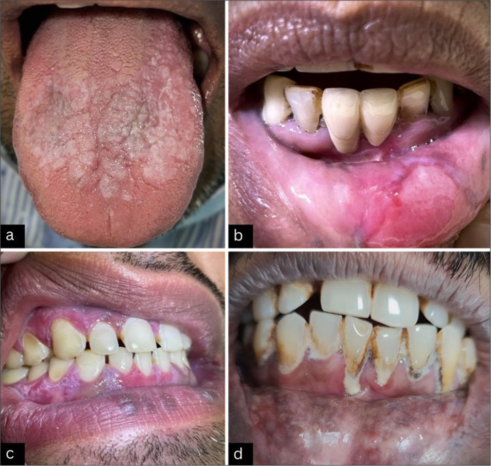
- Various presentations of mucosal lichen planus. (a): Multiple whitish irregular coalescing papules and plaques with the occasional reticulate arrangement over the tongue; (b): Well defined, pale to red superficial erosion with irregular intensive red edge and white hyperkeratotic periphery over labial mucosa (bi-color); (c): Whitish lacy pattern on gingiva; (d): Slightly atrophic grayish-black pigmentation over the upper lip and few micro-erosions with peripheral brownish to violaceous pigmentation and irregular white lacy network over labial mucosa.

- Dermoscopy of mucosal lichen planus. (a): Linear and radial Wickham striae (red circle) over a background of brown-black globules (black star) with linear and dotted vessels (black arrow) arranged irregularly over labial mucosa; (b): microerosion surrounded by white veil-like structure less area (black circle) with a diffused background of brown-black globules (black star); (c): diffused white to gray structure less area with occasional linear Wickham striae (red circle), bleeding spots, and hairpin vessels (black arrow) and white clods probably blunted lingual papillae (black circle) over the tongue; (d): Wickham’s striae and white to gray structure less area over gingival mucosa; (e): annular and radial Wickham’s striae (red circle) with multiple bleeding spots (black star); and (f): large, pale white to reddish erosion with few bleeding spots (black star), multiple linear vessels, intensive red edge (red arrow), and white veil-like structure less periphery (yellow star). Radial arrangement of Wickham’s striae (red circle) over lips.
DISCUSSION
Oral LP may occur in isolation (25%), or it can be part of mucocutaneous LP (30–77%).[1,2] Oral LP can affect buccal mucosa, glossal mucosa, gingiva, and lip mucosa, either in isolation or multiregional involvement.[3] We observed a predominant involvement of lip mucosa in our patients. WS is a pathognomic finding for the diagnosis of LP, which corresponds to compact orthokeratosis above wedge-shaped hypergranulosis on histopathology. On mucoscopy, various patterns of WS described are reticular, linear, radial streaming, globular, leaf-venation type, perpendicular, annular/circular, starry sky/white, and dots/starburst pattern.[2,4] In the present study, the radial pattern WS was most commonly observed. In addition, we also noticed modified WS patterns such as hyperkeratotic leukoplakia-like areas[5] and veil-like structureless gray-white to bluish-white areas and speckled-pearly areas[6] in our patients with lip and glossal LP, respectively. Further, bi-colored and tri-colored patterns, characterized by a combination of WS with bright red moist erosions and purple to brown colored plaques, respectively, have also been observed.[5,6] Vascular patterns, including dotted, linear, curvilinear, hairpin, or looped patterns have been described for both mucosal and cutaneous LP.[2,4,6,7] Linear pattern (n = 6) and dotted pattern (n = 4) was observed in our series. In the pigmentation pattern gray-black granules, globules, streaks of brown pigmentation, and violaceous to brown colored clods have been described, and it correlates with basal layer degeneration and pigment incontinence on histopathology.[2,4,6] In a study of 12 biopsy-confirmed cases of lip LP, Neema et al., additionally reported erosions (50%), bleeding spots (33%), and rosettes (four-dot sign) at the lip margin [Table 1].[7]
| Study | Neema et al.[7] (%) | *Rouai et al.[2] (%) | Our study (%) |
|---|---|---|---|
| Number of patients | 12 | 27 | 11 |
| Mucoscopic features | |||
| Wickham striae (n, %) (most common pattern) |
12 (100) (radial) |
24 (91) (reticular) |
11 (100) (radial and linear) |
| LLA or veil-like structureless zones (Modified Wickham striae) (n, %) |
0 | 5 (19) | 3 (27) |
| Vessels (most common pattern) |
11 (91) (linear) |
23 (88) (linear) |
9 (81) (linear) |
| Pigmented structures (most common pattern) |
12 (100) (Gray-black granules, globules) |
11 (41) (brown/blue globules) |
4 (36) (brown/black globules) |
| Erosions | 6 (50) | 17 (63) | 3 (27) |
| Scales | 12 (100) | 9 (34) | 0 |
| Bleeding spots | 4 (33) | 0 | 4 (36) |
| Rosettes | 4 (33.3) | 0 | 0 |
Despite the small sample size and challenges in accessing the palate and posterior buccal mucosa, mucoscopy establishes itself as a promising adjunct in diagnosing oral LP.
Innovative approaches, such as attaching disposable plastic tubes to universal serial bus (USB) dermatoscopes[8] or utilizing chalazion clamps[9] have greatly improved visualization of the anterior oral cavity. However, examining the posterior oral cavity still remains a challenge as traditional methods for posterior oral cavity examination limit adequate visualization. A structurally modified dermatoscope with features such as adjustable angles, retractable retractors, and longer but slender handle extensions specifically designed for navigating the intricacies of the oral cavity would be a valuable tool in addressing this limitation. The development of a dedicated oral dermatoscope holds the promise of revolutionizing mucoscopy for the entire oral cavity.
CONCLUSION
While the current literature on mucoscopy of oral LP is limited, this technique demonstrates potential in visualizing Wickham striae and other characteristic features, thereby, at times, obviating the need for biopsies. The enhanced visualization offered by mucoscopy may improve patient compliance and consequently treatment outcomes.
Ethical approval:
The Institutional Review Board approval is not required.
Declaration of patient consent:
The authors certify that they have obtained all appropriate patient consent.
Conflicts of interest:
There are no conflicts of interest.
Use of artificial intelligence (AI)-assisted technology for manuscript preparation:
The authors confirm that there was no use of artificial intelligence (AI)-assisted technology for assisting in the writing or editing of the manuscript and no images were manipulated using AI.
Financial support and sponsorship: Nil.
References
- Application of mucous membrane dermoscopy (mucoscopy) in diagnostics of benign oral lesions-literature review and preliminary observations from international dermoscopy society study. Dermatol Ther. 2021;34:e14478.
- [CrossRef] [PubMed] [Google Scholar]
- Dermoscopic features of mucosal lichen planus. Int J Dermatol. 2021;60:1368-72.
- [CrossRef] [PubMed] [Google Scholar]
- The clinical manifestations and treatment of oral lichen planus. Dermatol Clin. 2003;21:79-89.
- [CrossRef] [PubMed] [Google Scholar]
- Dermoscopy of oral mucosal lesions: Experience from a tertiary care center in North India and review of Literature. Indian Dermatol Online J. 2022;13:346-60.
- [CrossRef] [PubMed] [Google Scholar]
- Clinical diagnosis of oral erosive lichen planus by direct oral microscopy. Postepy Dermatol Alergol. 2014;31:222-8.
- [CrossRef] [PubMed] [Google Scholar]
- Case report: Dermoscopic features of oral lichen planus-the evolution of mucoscopy. F1000Res. 2018;7:284.
- [CrossRef] [PubMed] [Google Scholar]
- Dermoscopy of lip lichen planus-a descriptive study. Dermatol Pract Concept. 2020;10:e2020076.
- [CrossRef] [PubMed] [Google Scholar]
- Innovative modification of the USB dermatoscope for mucoscopy. J Am Acad Dermatol. 2018;78:e3-4.
- [CrossRef] [PubMed] [Google Scholar]
- Using a chalazion clamp to enhance dermoscopy of oral mucosal lesions. J Am Acad Dermatol. 2017;76:e91-2.
- [CrossRef] [PubMed] [Google Scholar]


