
Translate this page into:
Psoralen and ultraviolet -A therapy in folliculotropic mycosis fungoides: Long-term outcome in a young female patient – A 4-year follow-up
*Corresponding author: Twaseem Padiyath Mohamed, Department Dermatology, Amala Institute of Medical Sciences, Thrissur, Kerala, India. twaseem16@gmail.com
-
Received: ,
Accepted: ,
How to cite this article: Mohamed TP, Criton SP, Abraham UM. Psoralen and ultraviolet -A therapy in folliculotropic mycosis fungoides: Long-term outcome in a young female patient – A 4-year follow-up. J Skin Sex Transm Dis. doi: 10.25259/JSSTD_12_2025
Abstract
Folliculotropic mycosis fungoides (FMF) is a rare variant of cutaneous T-cell lymphoma characterized by folliculotropic lymphocytic infiltration, and its diagnosis is challenging due to the diverse clinical manifestations, especially in patients with atypical presentations and skin color. We report the case of a 28-year-old female with skin of color who presented with a hypopigmented, atrophic facial plaque, which was initially misdiagnosed and inadequately treated. Histopathology and immunohistochemistry confirmed the diagnosis of FMF, classified as stage 1A. Following the Dutch cutaneous lymphoma group guidelines, a treatment plan of 10 sessions of psoralen and ultraviolet-A therapy was implemented, leading to significant clinical and histopathological improvement. The patient has remained in remission for 4 years of follow-up without the need for further interventions. This case underscores the diagnostic complexities and therapeutic challenges associated with FMF, particularly in younger patients and underrepresented populations, and emphasizes the need for inclusive research to develop personalized treatment guidelines. It highlights the importance of early diagnosis, personalized therapy, and diversity in clinical research to optimize outcomes for patients with FMF.
Keywords
Folliculotropic mycosis fungoides
Psoralen and UV-A therapy
Skin of color

INTRODUCTION
Folliculotropic mycosis fungoides (FMF) is a rare variant of cutaneous T-cell lymphoma, constituting 10% of all cases of mycosis fungoides (MF) primarily reported among adult men and is rarely found in adolescents and children. In most cases, they present as follicular papules, acneform lesions, erythematous patches and plaque, alopecic patches with or without scarring, and nodular or prurigo-type lesions involving the face, neck, and upper trunk, unlike MF, which spares this area. Histopathologically, it is characterized by deep follicular and perifollicular infiltrates of lymphocytes sparing the epidermis.
The clinical course and response to treatment in FMF can vary significantly, with limited evidence on long-term outcomes, especially in younger patients and with darker skin types. Here, we report the case of a 28-year-old female who presented with a hypopigmented facial plaque diagnosed with FMF. The case highlights the diagnostic complexities and therapeutic considerations, particularly without robust, diverse clinical data. This report also emphasizes the significance of inclusivity in clinical research and guideline development to improve outcomes for diverse populations.
CASE REPORT
A 28-year-old female with no comorbidities presented to the dermatology outpatient department with a hypopigmented lesion on the right side of her face, which was associated with occasional itching for 6 months [Figure 1]. Initially, she consulted a physician and was given topical mid-potent steroid and oral antihistamine for 1 month, following which the lesion subsided, but soon reappeared after stopping the medication with more scaling and gained size over 6 months. A dermatological examination showed a hypopigmented atrophic plaque with follicular papules, loss of hair, and fine scaling in the temporomandibular area. With the clinical differential diagnosis of borderline leprosy, idiopathic follicular mucinosis, atypical lichen plano pilaris, and FMF, a biopsy was taken from the facial lesion, which showed accumulation of mucin in the follicular epithelium and lymphocytic infiltrate surrounding the hair follicle [Figure 2]. Immunohistochemistry showed CD3 + [Figure 3] CD4 + CD8 + CD20 reactive cell + and loss of expression of CD7 [Figure 4], CD5. CD30, S100, and CD68 were also negative. These findings were consistent with FMF. Blood reports were within normal limits. Computed tomography scanning showed an ill-defined ground-glass nodule in the left upper lobe of the lung, which was suggestive of infective foci and was treated with azithromycin. Thus, the diagnosis was staged as FMF-1A [Table 1].[1]
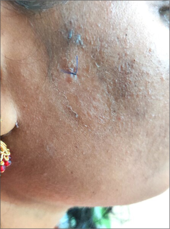
- Hypopigmented atrophic plaque on the face.

- Follicular mucinosis and lymphocytic infiltration on the histopathology-red arrow (hematoxylin and eosin staining, ×10 magnification).

- Immunohistochemistry showing CD 3 positivity (×10 magnification).

- Immunohistochemistry showing loss of expression of CD7 (×10 magnification).
| Stage | T | N | M | Description |
|---|---|---|---|---|
| I A | T1 | N0 | M0 | Localized patch/plaque <10% body surface area |
| I B | T2 | N0 | M0 | Diffuse patch/plaque >10% body surface area |
| II A | T1-2 | N1 | M0 | Patch/plaque and lymphadenopathy |
| II B | T3 | N0-1 | M0 | Tumor, lymphadenopathy |
| III | T4 | N0-1 | M0 | Erythroderma, lymphadenopathy |
T: Tumor, N: Node, M: Metastasis
The case was discussed in detail in our tumor board, and following recent guidelines from the Dutch cutaneous lymphoma group, a treatment plan was devised. Psoralen and ultraviolet-A therapy (PUVA) were initiated, starting at 2 joules and escalating to 9.5 joules. Remarkably, after 10 sessions of PUVA therapy on alternate days, there was clinical improvement, and a repeat biopsy showed a significant decrease in T-cell infiltration [Figure 5]. The patient has since been under follow-up for the past 4 years, with no recurrence and no requirement for further interventions such as radiotherapy or retinoid therapy.

- After 10 sessions of psoralen and ultraviolet-A treatment.
DISCUSSION
The term “FMF” was used in 1985 by Kim as he narrated a biopsy specimen that depicted perifollicular and follicular infiltrate of atypical lymphocytes sparing the epidermis.[2] The incidence of FMF is 0.64/100000 with a male-to-female ratio of 2–5:1. A recent study has found the 5-year survival of FMF to be 62% in the early stage and 40% in advanced stage.[3]
The hypopigmented plaque observed in the above case is more typical of classical MF rather than FMF. However, a similar presentation was reported in a case series of four FMF cases from India.[4] Therefore, this might be a form of presentation in FMF cases in skin of color types. In a case series by Baykal et al., several other atypical presentations of FMF identified were rosacea-like lesions, lichen spinulosus-like lesions in association with hypopigmentation and alopecia, lupus tumidus-like lesions, excoriations, and asymptomatic dome-shaped lesions with mucin.[5]
Treatments suggested for FMF are phototherapy, which includes PUVA therapy and UV-B therapy, interferon or retinoid, radiotherapy, or total surface electron beam therapy, depending on the disease subtype. Tailoring the treatment protocol was difficult in this case as there were not many reports on long-term treatment responses in young female patients of skin color. We adopted Dutch cutaneous lymphoma group guidelines based on retrospective analysis of the Dutch cutaneous lymphoma registry due to the lack of large-scale studies on the Asian population.[6] Furthermore, there are no studies that have assessed if the treatment outcomes are similar for the younger age group.
The tumor board decided on a non-aggressive treatment of giving 10 sessions of PUVA, to which the patient responded clinically and histopathologically. This report throws light on the difficulty in treatment decisions due to limited data on the established treatment guidelines involving a diverse population. Moreover, the published case reports and case series involving the underrepresented population had limited information on the long-term follow-up and treatment response.
This report highlights the need for maintaining registries for underrepresented ethnicities and advocates for concerted efforts to promote inclusivity and diversity in clinical research and guideline development.
CONCLUSION
This case highlights the diagnostic and therapeutic challenges of FMF in a young female with skin of color, presenting atypically with a hypopigmented plaque. Effective management using PUVA therapy led to sustained remission, emphasizing the need for inclusive research and personalized guidelines to address variations in disease presentation and treatment response across diverse populations.
Ethical approval
Institutional Review Board approval is not required.
Declaration of patient consent
The authors certify that they have obtained all appropriate patient consent.
Conflicts of interest
There are no conflicts of interest.
Use of artificial intelligence (AI)-assisted technology for manuscript preparation
The authors confirm that there was no use of artificial intelligence (AI)-assisted technology for assisting in the writing or editing of the manuscript and no images were manipulated using AI.
Financial support and sponsorship: Nil.
References
- Phototherapy for mycosis fungoides. Indian J Dermatol Venereol Leprol. 2015;81:124-35.
- [CrossRef] [PubMed] [Google Scholar]
- Follicular mycosis fungoides. Am J Dermatopathol. 1985;7:300-1.
- [CrossRef] [PubMed] [Google Scholar]
- Folliculotropic mycosis fungoides in a Latin American hospital: Survival analysis. Actas Dermosifiliogr. 2022;113:930-7.
- [CrossRef] [PubMed] [Google Scholar]
- Follicular mycosis fungoides-A report of four Indian cases. Indian J Med Paediatr Oncol. 2009;30:108-12.
- [CrossRef] [PubMed] [Google Scholar]
- Underrecognized clinical features of folliculotropic mycosis fungoides: A large clinical series. J Dtsch Dermatol Ges. 2017;15:289-99.
- [CrossRef] [Google Scholar]
- Recommendations for treatment in folliculotropic mycosis fungoides: Report of the Dutch cutaneous lymphoma group. Br J Dermatol. 2017;177:223-8.
- [CrossRef] [PubMed] [Google Scholar]


