
Translate this page into:
Quiz questions from pediatric dermatology
*Corresponding author: Aravind Sivakumar, Department of Dermatology, Jawaharlal Institute of Postgraduate Medical Education and Research, Gorimedu, Pondicherry, India. aravinddermat@gmail.com
-
Received: ,
Accepted: ,
How to cite this article: Sivakumar A. Quiz questions from pediatric dermatology. J Skin Sex Transm Dis 2024:6:71-6. doi: 10.25259/JSSTD_34_2023.
-
A 4-day-old neonate born out of a normal vaginal delivery presented with multiple pus-filled lesions all over the body since day 2 of life. It was not associated with any fever or systemic symptoms. On examination, there were multiple discrete pustules seen mainly over the trunk and extremities, surrounded by blotchy erythema [Figure 1]. Giemsa-stained preparation from the pustules showed numerous eosinophils. What is the diagnosis?
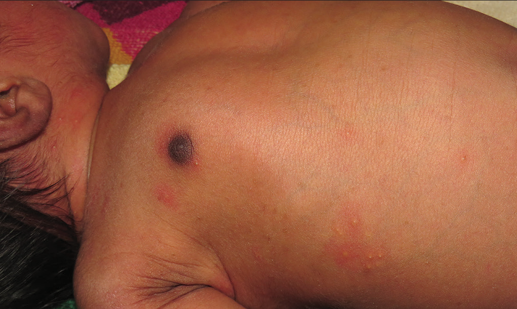 Figure 1:
- Discrete pustules surrounded by blotchy erythema over the trunk.
A 6-month-old male child presented with a band of alopecia encircling the parieto-occipital scalp. History revealed that the child was born of a prolonged vaginal delivery and had a large swelling of the scalp at birth. No local injuries or history of topical application was noted. What is the diagnosis?
A 12-year-old child with sparse hair growth complained of generalized decreased sweating. Examination revealed the presence of multiple fine pits along the dorsum of both forearms along with nodular lesions and milia over the face. Biopsy from the nodules was suggestive of basal cell carcinoma. What is the diagnosis?
-
A newborn baby presented with erosions over the scalp since birth [Figure 2]. On examination, there was a fine network of bluish atrophic patches over the acral areas which persisted even after warming, along with short distal phalanges of the fingers. What is the diagnosis?
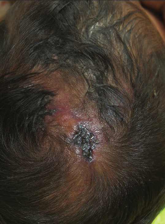 Figure 2:
- Well-defined ulcer of size 2 × 3 cm over the vertex of the scalp along with alopecia.
An 8-month-old infant presented with complaints of generalized exfoliation along with fever for 5 days. On examination, the child was irritable, febrile and there were crusted erosions with erythema over the periorificial and flexural regions, with areas of skin peeling. The Nikolsky sign was positive and the mucosal examination was normal. What is the diagnosis?
A 6-year-old child presented with a port wine stain involving the right side of the face and upper trunk, along with a persistent Mongolian spot on the lower back and limbs. There was history of seizure disorder and glaucoma. Imaging of the central nervous system (CNS) revealed evidence of cortical atrophy with dilated tortuous vessels. What is the diagnosis?
An 11-year-old boy presented with complaints of itchy oozy lesions over both lower limbs since 4 years of age. He also suffered from recurrent bouts of pneumonia, otitis media, sinusitis, and bronchial asthma. He claims that there is excessive bleeding from the skin on trivial trauma or scratching. There were two episodes of melena in the past. What is the diagnosis?
An 8-year-old child presented with multiple slow-growing painless nodules over the face for 3 years. Examination revealed a few well-defined firm- to- hard nodules of varying sizes over the face. Biopsy showed the presence of well-defined nodules composed of basaloid cells in the periphery and eosinophilic anucleate “ghost” cells in the center with areas of calcification. What are the systemic associations of this lesion?
-
A 5-year-old male child presented with complaints of lesions over the right side of the trunk since birth which were gradually increasing in size. Examination revealed the presence of linear hyperkeratotic papules in a Blashkoid distribution over the right side of the trunk, arm, and forearm up to the fingers [Figure 3]. A biopsy of the lesion revealed epidermolytic hyperkeratosis. What disease can be transmitted to further generations?
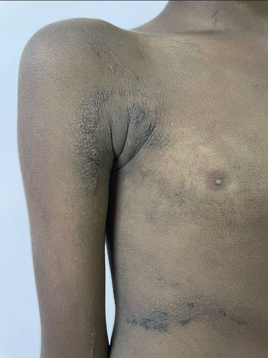 Figure 3:
- Well-defined hyperkeratotic blaschkoid plaque present over the trunk and the right upper extremity.
Which hormonal abnormality can be observed in cases of Langerhans cell histiocytosis in children?
Multiple lentigines with blue nevi arising in a background of endocrine dysfunction and myxoma of the heart are known as?
Large segmental hemangiomas of the face need screening for PHACES syndrome, what does “S” stand for in PHACES?
Multiple venous malformations of the skin and gastrointestinal (GI) intestinal tract caused by a mutation in the TIE2 gene are referred to as?
Which subcutaneous implant approved by Food and Drug Administration in 2019 is used for erythropoietic protoporphyria?
Which oral agent is approved in 2020 for prophylaxis in children above 12 years with hereditary angioneurotic edema (HAE)?
Bilateral eyelid swelling in a child with fever, maculopapular rash, tonsillitis, hepatosplenomegaly, and atypical lymphocytosis is referred to as?
-
Match the following disorders of cornification:
a. Jordans anomaly,
hepatosplenomegaly,
myopathy:Netherton
syndromeb. Cobblestone skin lesions,
sensorineural hearing loss,
keratitis:Refsum
syndromec. Sectorial cataracts, stippled
calcification, feathery mosaic
lesions:Chanarin
Dorfman
syndromed. Ichthyosis linearis
circumflexa, bamboo hair,
trichorrhexis invaginata:KID syndrome e. Cerebellar ataxia, peripheral
neuropathy, retinitis
pigmentosa:Conradi
Hunermann
Happle syndrome Epidermolysis bullosa with muscular dystrophy occurs due to a mutation in genes?
-
Diffuse palmoplantar keratoderma along with periodontitis [Figure 4] and recurrent infections is referred to as?
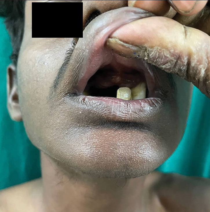 Figure 4:
- Periodontitis causing premature loss of teeth.
Woolly hair with palmoplantar keratoderma developing right ventricular cardiomyopathy occur due to mutation in?
In what syndrome does symmetrical areas of depigmentation with white forelock along with deafness and heterochromia iridis occur
Precocious puberty (PP), café au lait macules along with polyostotic fibrous dysplasia (FD) constitute?
Neurofibromatosis-like condition which has macrocephaly, café au lait macules, and freckling but doesn’t have neurofibroma, lisch nodules, or glioma occurs due to mutation in?
An eye examination is mandatory in cases of early onset lymphedema in childhood to exclude?
Which condition that occurs in infants can mimic Henoch Schonlein purpura with purpura, edema, and arthralgia?
A child with multiple onset elastomas of the skin is likely to have which imaging findings in the bones?
Reticulate pigmentation along with premalignant oral leukoplakia and nail dystrophy is seen in which condition?
Which inherited condition with photosensitivity presents with bird head facies, mickey mouse ears, mental retardation along with salt and pepper retinal degeneration?
Which chromosomal aneuploidy can have features of recurrent leg ulcers, prone to lupus erythematosus, and in which male neonates survive X- linked dominant conditions?
Urine examination in a child with acroparesthesias, multiple angiokeratomas, and corneal opacities is likely to show what finding?
Answers:
-
Erythema toxicum neonatorum
It is a misnomer, being a transient benign neonatal dermatosis, self-resolving within the first few weeks, and occurs usually at day 2–3 days after birth. Etiology remains unclear, with commensal microbial organisms being implicated in the innate immune response.[1]
-
Halo scalp ring
It occurs because of prolonged labor that results in caput succedaneum with pressure necrosis. It presents as a band of alopecia over the vertex bordering the caput and can occur during birth or a few days after birth. Regrowth of hair usually happens which is usually non-scarring in few months to years, however, scarring can occur in deep seated injuries due to necrotic or hemorrhagic caput succedaneum. It needs to be differentiated from other causes of alopecia such as neonatal occipital alopecia, pressure, and trauma induced causes.[2]
-
Bazex Dupre'Christol syndrome
It is a rare X-linked dominant genodermatosis characterized clinically by the triad of hypotrichosis, basal cell carcinoma and follicular atrophoderma. Other findings include milia, hypohidrosis, trichoepithelioma, and ichthyosis. It should not be confused with acrokeratosis paraneoplastica of Bazex which is a paraneoplastic phenomenon.[3]
-
Adams Oliver syndrome
It is a rare multiple malformation syndrome characterized by aplasia cutis congenita along with transverse terminal limb defects. Furthermore, it can present with cutis marmorata telangiectasia congenita, congenital heart defects, and CNS anomalies.[4]
-
Staphylococcal scalded skin syndrome (SSSS)
SSSS also known as Ritter’s disease is a dermatological emergency characterized by diffuse blistering and denudation of skin with positive Nikolsky and sparing the mucosal surfaces. It occurs due to the production of exotoxin by staphylococcus aureus that cleaves desmoglein 1 in the skin.[5]
-
Phakomatosis pigmentovascularis(PPV)
PPV is a mosaic disorder of melanocytes and vasomotor nerves due to sporadic mutations in GNAQ/GNA11. This subtype can be classified as type 2 or cesioflammea with nevus flammeus along with dermal melanocytosis and type 2B due to systemic involvement.[6]
-
Wiskott-Aldrich syndrome(WAS)
WAS is a rare X-linked recessive disorder that presents with eczema, bleeding manifestations due to microthrombocytopenia and recurrent infections due to mutations in WAS gene with defective WASp protein.[7]
Gardner syndrome, MYH-associated polyposis, Rubinstein-Taybi syndrome, Sotos syndrome, myotonic dystrophy, gliomatosis cerebri, and Turner syndrome. These are multiple pilomatricoma associated syndromes.[8]
Epidermolytic ichthyosis/bullous ichthyosiform erythroderma. Parents who have epidermal nevi which is epidermolytic in histology may transmit to their offspring due to presence of germline mosaicism in keratin encoding genes mainly KRT1/KRT10.[9]
Diabetes insipidus or anterior hypopituitarism can be observed in cases of pituitary involvement in Langerhans cell histiocytosis. It is a risk factor for neurodegeneration occurring as a late complication.[10]
-
Carney complex/LAMB/NAME syndrome
It is an autosomal dominant condition due to mutation in PRKAR1A characterized by the presence of spotty skin pigmentation or lentigines, endocrine overactivity, and myxomas. Cutaneous manifestations can include blue nevi, compound or junctional melanocytic nevi and cutaneous myxomas.[11]
-
Sternal deformities
PHACES syndrome comprises posterior fossa malformations, segmental hemangioma of the face, arterial anomalies, coarctation of the aorta/cardiac defects, and eye abnormalities. Sternal abnormalities was added later, which include sternal cleft and supraumbilical raphae.[12]
-
Bean syndrome/Blue rubber bleb nevus syndrome
Characterized by multiple venous malformations of the skin, soft tissue and GI tract. Needs evaluation for GI involvement as these patients can have chronic bleeding manifestations and anemia needing frequent blood transfusions or surgery.[13]
-
Afamelanotide
As an alpha MSH analogue it decreases symptoms in patients with erythropoietic protoporphyria by causing increase in eumelanogenesis without sun exposure. Other uses are for vitiligo, solar urticaria and Hailey Hailey disease.[14]
-
Berotralstat
It is an oral plasma kallikrein inhibitor taken once daily for long-term prophylaxis of HAE in children above 12 years of age.[15]
-
Hoagland sign
Occurs in infectious mononucleosis caused by Ebstein Barr virus infection.[16]
-
Match the following disorders of cornification: [17]
Jordans anomaly, hepatosplenomegaly, myopathy: Chanarin Dorfman syndrome
Cobblestone lesions, Sensorineural hearing loss, keratitis: KID syndrome
Sectoral cataracts, Stippled calcification, feathery mosaic lesions: Conradi Hünermann Happle syndrome
Ichthyosis linearis circumflexa, Bamboo hair, trichorrhexis invaginata: Netherton syndrome
Cerebellar ataxia, peripheral neuropathy, retinitis pigmentosa: Refsum syndrome
Plectin mutation[18]
-
Papillon Lefèvre syndrome
It as a autosomal recessive palmoplantar keratoderma with periodontitis leading to premature loss of deciduous teeth and recurrent skin infections due to mutation in Cathepsin C.[19]
-
Naxos syndrome
Occurs due to mutation in plakoglobin gene, manifests with woolly hair, palmoplantar keratoderma along with arrhythmogenic right ventricular cardiomyopathy.[20]
-
Waardenburg syndrome
Characterized by depigmented patches of the skin, white forelock, heterochromia irides, sensorineural hearing loss. In addition patients can have dystopia canthorum, musculoskeletal abnormalities, and Hirschprung’s disease.[21]
-
McCune Albright syndrome (MAS)
MAS is a sporadic mosaic disorder caused by mutation in GNAS1 characterized by the triad of monostotic/polyostotic FD, café au lait skin pigmentation, and hyperfunctioning endocrinopathies, including gonadotropin-independent PP, thyrotoxicosis, growth hormone excess, hyperprolactinemia, or neonatal hypercortisolism.[22]
-
Legius syndrome
It is milder variant of neurofibromatosis type 1 (NF-1) caused by SPRED-1 mutation characterized by macrocephaly and cafe au lait with or without freckling. Other signs of NF-1 are absent.[23]
-
Lymphedema Distichiasis syndrome
Double rows of eyelashes are referred to as distichiasis, which can be associated with onset of lymphedema in late childhood or puberty and venous insufficiency caused due to FOXC2 mutation. Other anomalies that can be associated include congenital heart disease, ptosis, cleft lip/palate, and spinal extradural cysts.[24]
-
Acute hemorrhagic edema of infancy
Also referred to as Finkelstein disease or Seidlmayer purpura, it is a self-limited benign condition of infancy presenting as fever, targetoid purpura, edema, and arthralgia.[25]
-
Osteopoikilosis
Buschke-Ollendorf syndrome is an autosomal recessive condition due to mutation in LEMD3/MAN1 characterized by multiple collagen and elastin nevi referred to as dermatofibrosis lenticularis disseminata. Imaging reveals presence of bone islands called osteopoikilosis.[26]
-
Dyskeratosis congenital
It is an inherited bone marrow failure syndrome commonly due to mutation in dyskerin or TERT characterized by the triad of nail dystrophy, reticulate skin pigmentation along with oral leukoplakia, can predispose to aplastic anemia, myelodysplastic syndrome, leukemia, and other solid organ malignancies.[27]
-
Cockayne syndrome
It belongs to the group of nucleotide excision repair disorders characterized by their prominent facial features, photosensitivity, mental retardation, microcephaly, hearing loss, and retinal degeneration.[28]
Klinefelter syndrome[29]
-
Maltese cross
This is Fabry disease, due to deficiency of alpha galactosidase A, which presents with angiokeratoma corporis diffusum, acroparesthesia, hypohidrosis, and corneal opacities. Urine examination reveals Maltese cross globules under polarizing microscope.[30]
Ethical approval
The Institutional Review Board approval is not required.
Declaration of patient consent
The authors certify that they have obtained all appropriate patient consent.
Conflicts of interest
There are no conflicts of interest.
Use of artificial intelligence (AI)-assisted technology for manuscript preparation
The author(s) confirms that there was no use of Artificial Intelligence (AI)-Assisted Technology for assisting in the writing or editing of the manuscript and no images were manipulated using the AI.
Financial support and sponsorship
Nil.
References
- Erythema toxicum neonatorum is an innate immune response to commensal microbes penetrated into the skin of the newborn infant. Pediatr Res. 2005;58:613-6.
- [CrossRef] [PubMed] [Google Scholar]
- Halo scalp ring: A case series and review of the literature. Arch Pediatr Adolesc Med. 2002;156:188-90.
- [CrossRef] [PubMed] [Google Scholar]
- Bazex-Dupré-Christol syndrome: Review of clinical and molecular aspects. Int J Dermatol. 2018;57:1102-6.
- [CrossRef] [PubMed] [Google Scholar]
- Adams-Oliver syndrome review of the literature: Refining the diagnostic phenotype. Am J Med Genet A. 2017;173:790-800.
- [CrossRef] [PubMed] [Google Scholar]
- Staphylococcal scalded skin syndrome: An epidemiological and clinical review of 84 cases. Pediatr Dermatol. 2021;38:149-53.
- [CrossRef] [PubMed] [Google Scholar]
- Phacomatosis pigmentovascularis revisited and reclassified. Arch Dermatol. 2005;141:385-8.
- [CrossRef] [PubMed] [Google Scholar]
- Wiskott-Aldrich syndrome: A comprehensive review. Ann N Y Acad Sci. 2013;1285:26-43.
- [CrossRef] [PubMed] [Google Scholar]
- Pilomatrixoma: A comprehensive review of the literature. Am J Dermatopathol. 2018;40:631-41.
- [CrossRef] [PubMed] [Google Scholar]
- Epidermolytic ichthyosis in a child and systematized epidermolytic nevi in the mosaic parent associated with a KRT1 variant. Eur J Med Genet. 2021;64:104324.
- [CrossRef] [PubMed] [Google Scholar]
- Hand Schuller Christian disease. Indian J Med Paediatr Oncol. 2011;32:183-4.
- [CrossRef] [PubMed] [Google Scholar]
- PHACE syndrome: Clinical manifestations, diagnostic criteria, and management. An Bras Dermatol. 2018;93:405-11.
- [CrossRef] [PubMed] [Google Scholar]
- Blue rubber bleb nevus syndrome: Surgical eradication of gastrointestinal bleeding. Ann Surg. 2005;241:523-8.
- [CrossRef] [PubMed] [Google Scholar]
- Afamelanotide for prevention of phototoxicity in erythropoietic protoporphyria. Expert Rev Clin Pharmacol. 2021;14:151-60.
- [CrossRef] [PubMed] [Google Scholar]
- Berotralstat (BCX7353) is a novel oral prophylactic treatment for hereditary angioedema: Review of phase II and III studies. Allergy Asthma Proc. 2021;42:274-82.
- [CrossRef] [PubMed] [Google Scholar]
- Hoagland sign in infectious mononucleosis. BMJ Case Rep. 2022;15:e252839.
- [CrossRef] [PubMed] [Google Scholar]
- Revised nomenclature and classification of inherited ichthyoses: Results of the First Ichthyosis Consensus Conference in Sorèze 2009. J Am Acad Dermatol. 2010;63:607-41.
- [CrossRef] [PubMed] [Google Scholar]
- Epidermolysis bullosa simplex with muscular dystrophy. Review of the literature and a case report. J Dermatol Case Rep. 2016;10:39.
- [CrossRef] [PubMed] [Google Scholar]
- Naxos disease: Cardiocutaneous syndrome due to cell adhesion defect. Orphanet J Rare Dis. 2006;1:4.
- [CrossRef] [PubMed] [Google Scholar]
- Review and update of mutations causing Waardenburg syndrome. Hum Mutat. 2010;31:391-406.
- [CrossRef] [PubMed] [Google Scholar]
- Clinical characteristics, and management of patients with McCune-Albright syndrome with GH excess and precocious puberty: A case series and literature review. Front Endocrinol. 2021;12:672394.
- [CrossRef] [PubMed] [Google Scholar]
- Review and update of SPRED1 mutations causing Legius syndrome. Hum Mutat. 2012;33:1538-46.
- [CrossRef] [PubMed] [Google Scholar]
- Lymphedema-distichiasis syndrome: A distinct type of primary lymphedema caused by mutations in the FOXC2 gene. Int J Dermatol. 2008;47:52-5.
- [CrossRef] [PubMed] [Google Scholar]
- Acute hemorrhagic edema of infancy: A worrisome presentation, but benign course. Clin Cosmet Investig Dermatol. 2013;6:197-9.
- [CrossRef] [PubMed] [Google Scholar]
- The Buschke-Ollendorff syndrome: A case report of simultaneous osteo-cutaneous malformations in the hand. BMC Res Notes. 2016;9:294.
- [CrossRef] [PubMed] [Google Scholar]
- Dyskeratosis congenita. Hematol Oncol Clin North Am. 2009;23:215-31.
- [CrossRef] [PubMed] [Google Scholar]
- Dermatologic findings in 16 patients with cockayne syndrome and Cerebro-Oculo-Facial-Skeletal syndrome. JAMA Dermatol. 2013;149:1414-8.
- [CrossRef] [PubMed] [Google Scholar]
- Leg ulcers in Klinefelter's syndrome--further evidence for an involvement of plasminogen activator inhibitor-1. Br J Dermatol. 1997;136:341-4.
- [CrossRef] [PubMed] [Google Scholar]


