Translate this page into:
Ultraviolet-induced fluorescence trichoscopy of trichobacteriosis axillaris
*Corresponding author: Deepika Yadav, Department of Dermatology and Venereology, Maulana Azad Medical College, New Delhi, India. deepikayadav18.90@gmail.com
-
Received: ,
Accepted: ,
How to cite this article: Gaurav V, Modi V, Yadav D. Ultraviolet-induced fluorescence trichoscopy of trichobacteriosis axillaris. J Skin Sex Transm Dis. 2024;6:217-8. doi: 10.25259/JSSTD_21_2024
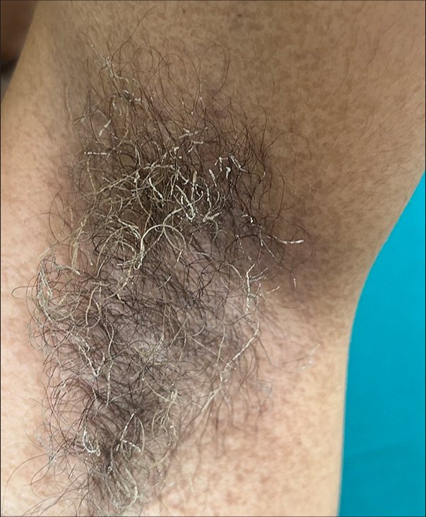
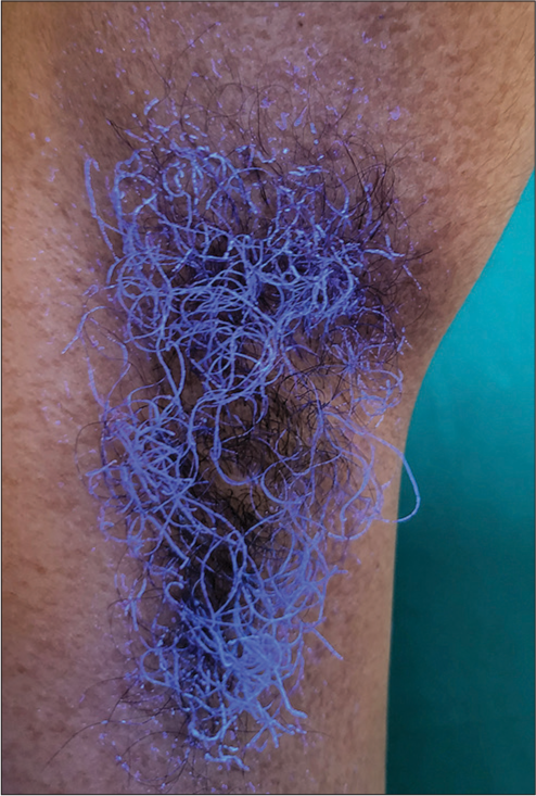
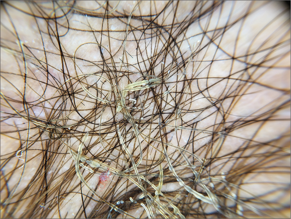
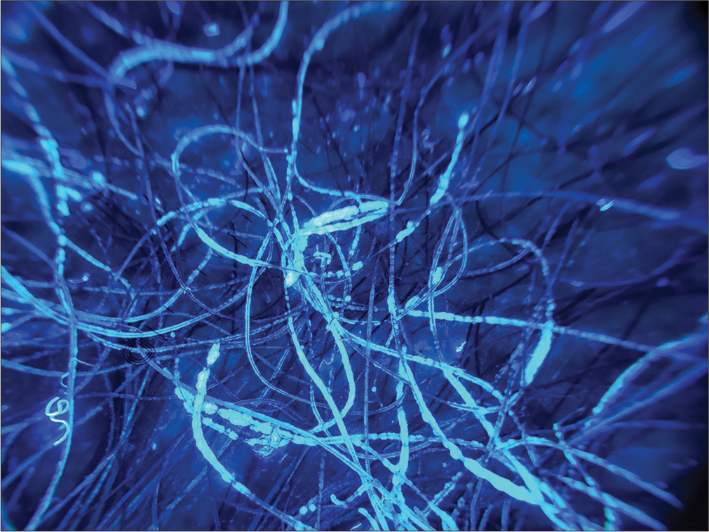
A 23-year-old female presented with complaint of malodour from bilateral axillae for two years, which worsened during summers. Examination revealed firmly adherent, yellow-white concretions along multiple hair shafts in both axillae associated with an offensive smell [Figure 1]. These concretions showed bluish fluorescence under Wood’s lamp [Figure 2]. Polarized trichoscopy showed pale-yellow, cotton-like waxy structures forming sheaths, nodules, and concretions along the hair shafts [Figure 3], which showed bluish fluorescence on ultraviolet-induced fluorescence trichoscopy [Figure 4]. Microscopic examination of 10% potassium hydroxide mount revealed irregular cottony concretions along hair shafts. She was diagnosed with trichobacteriosis axillaris (trichomycosis axillaris) based on the above findings. The patient was advised to shave her axillary hair and prescribed clindamycin phosphate 1% gel for twice daily topical application resulting in remission within two weeks.

- Firmly adherent, yellow-white concretions along multiple hair shafts over right axilla.

- Bluish fluorescence under Wood’s lamp.

- Polarized trichoscopy showing pale-yellow, cotton-like waxy structures forming sheaths, nodules, and concretions along the hair shafts (DermLite DL5, ×20).

- Ultraviolet-induced fluorescence trichoscopy showing bluish fluorescent concretions (DermLite DL5, ×20).
Ethical approval
The Institutional Review Board approval is not required.
Declaration of patient consent
The authors certify that they have obtained all appropriate patient consent.
Conflicts of interest
There are no conflicts of interest.
Use of artificial intelligence (AI)-assisted technology for manuscript preparation
The authors confirm that there was no use of artificial intelligence (AI)-assisted technology for assisting in the writing or editing of the manuscript and no images were manipulated using AI.
Financial support and sponsorship
Nil.


