
Translate this page into:
Erythema multiforme-like rash as a manifestation of multisystem inflammatory syndrome in children
*Corresponding author: Sandhya George, Additional professor, Department of Dermatology and Venereology, Government Medical College, Manjeri, Hospital Road, Malappuram, Kerala, India. drsandhyageorge@gmail.com
-
Received: ,
Accepted: ,
How to cite this article: Sindhu CB, Francis B, George S, Reena Mariyath OK, Peethambaran G, Shams S. Erythema multiforme-like rash as a manifestation of multisystem inflammatory syndrome in children. J Skin Sex Transm Dis 2021;3:181-3.
Abstract
Multisystem inflammatory syndrome in children (MIS-C) is a rare and serious manifestation of coronavirus disease 19 (COVID-19) infection. Skin lesions occur in more than half the cases of MIS-C. We report a 57-dayold female baby who presented with features of MIS-C with skin lesions suggestive of erythema multiforme. Her condition improved rapidly with systemic steroids and intravenous immunoglobulin G.
Keywords
Coronavirus disease-19 infection
Multisystem inflammatory syndrome
Children
Erythema multiforme
Skin lesion

INTRODUCTION
Coronavirus disease-19 (COVID-19), a viral infection, caused by severe acute respiratory syndrome corona virus-2 (SARS-CoV-2) affects all age groups. The infection usually manifests with less severity in children. Recently, a severe multisystem inflammatory syndrome has been reported in individuals <21 years of age.[1] The frequency of skin involvement is more than 50% in multisystem inflammatory syndrome in children (MIS-C) and the lesions vary in presentation.[2] Erythema multiforme (EM) is a rare presentation of MIS-C, with only a few reported cases.[3]
CASE REPORT
A 57-day-old female child was brought to Government Medical College, Manjeri, with a skin rash of 10 days and fever of 1 week duration. She also had cough and breathlessness. The baby and her mother tested positive for SARS-CoV-2 by real-time reverse-transcriptase polymerase chain reaction (RT-PCR).
Baby weighed 4.1 kg. Her pulse and respiratory rates were 160/minute and 56/minute, respectively. At the time of admission, she was hemodynamically stable and afebrile. Oxygen saturation was 96%. Dermatological examination showed lesions of different morphology ranging in size from 5 mm to 4.5 cm and included erythematous plaques, papules and papulovesicles. Larger plaques were seen mainly over the face and resembled target lesions with central cyanotic hue, surrounding pallor and peripheral erythema [Figure 1]. On the 2nd day, the lesions increased in size [Figure 2]. Some of the lesions differed from the typical target lesions by being more than 3 cm in size. Papulovesicles were mainly seen on the limbs. Mucosae were spared.
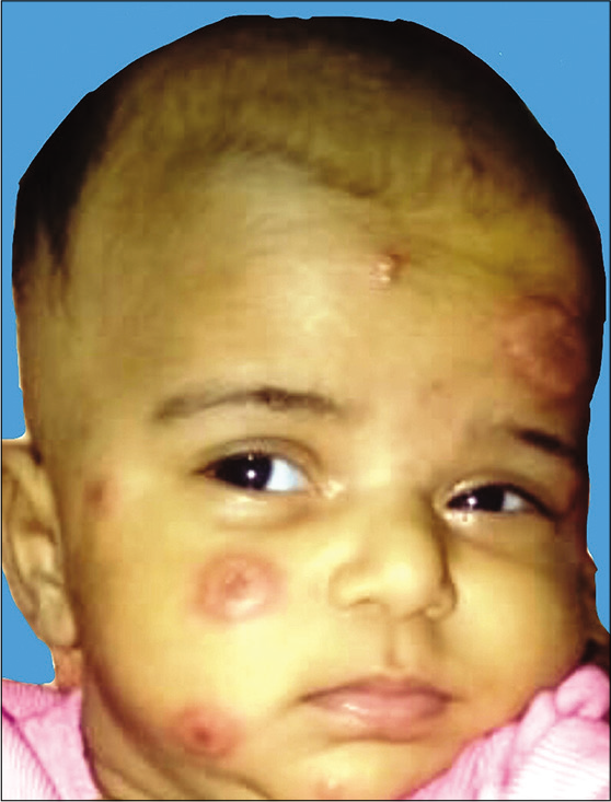
- Erythematous plaque with central cyanotic hue on the face of an infant with multisystem inflammatory syndrome in children (1st day of admission).
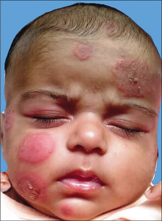
- The same child shown in Figure 1 on the 2nd day of admission. Note the increase in size of the rash.
Hemoglobin level which was 9.5 g% at admission dropped to 8.6 g% in 2 days, while platelet count rose from 5 lakh cells/ mm3 to 9 lakh cells/mm3. Total leukocyte count was 35,750 cells/mm3. Erythrocyte sedimentation rate and C-reactive protein were 32 mm/hour and 144 mg%, respectively. The patient required ionotropic support as she developed hypotension. Serum ferritin increased from 923 ng/ml to 2000 ng/ml in 2 days. Chest X-ray, echocardiogram, serum troponin, serum fibrinogen, D-dimer, and renal and liver function tests were normal. Blood culture was sterile.
Considering the target lesions, systemic involvement, and investigation results, a diagnosis of EM-like rash secondary to MIS-C was made. Our patient completed the case definition of MIS-C, as she had fever of more than 3 days, rash, systemic involvement, elevated markers of inflammation, and positive RT-PCR with other microbial causes of inflammation ruled out. The baby was referred to Government Medical College, Kozhikode, where she received intravenous methyl prednisolone 20 mg/kg/day for 3 days and intravenous immunoglobulin G 400 mg/kg/day for 2 days along with antibiotics. The child showed good response to treatment. She did not develop respiratory distress at any time during the course of the disease.
Skin lesions started subsiding by the 3rd day. Prednisolone 10 mg per orally (for 10 days) substituted the parenteral methyl prednisolone after 3 days and low-dose aspirin (10 mg/kg) was added to the treatment regimen. Repeat echocardiography was normal and she was discharged on the 10th day of admission. Currently, 1 month after the discharge, the child is not on any drugs and remains asymptomatic.
DISCUSSION
MIS-C during the course of COVID-19 infection has been reported from different parts of the world.[1-3] MIS-C manifests with varying features such as fever, skin rash, shock, cardiac dysfunction, abdominal pain and elevated inflammatory markers (CRP, ferritin, D-dimer, and interleukin-6).[2]
The case definition of MIS-C by Centers for Disease Control and Prevention includes six criteria: Serious illness leading to hospitalization, age <21 years, fever (body temperature >38.0°C) or report of subjective feeling of fever lasting at least 24 hours, laboratory evidence of inflammation, multisystem organ involvement and laboratory-confirmed SARS-CoV-2 infection, or an epidemiologic link to a person with COVID-19.[1]
In a typical case of MIS-C, children show signs/symptoms 3–4 weeks after COVID-19 infection. Some may progress rapidly to shock and cardiorespiratory failure. With prompt recognition and medical attention, most children survive, but the long-term outcomes are presently unknown.[4]
In a case series of 58 children with MIS-C, the documented findings were fever, myocardial injury, shock, and coronary artery aneurysm. Majority had abdominal pain/diarrhea as the presenting symptom.[5] Most of the affected children were treated with intravenous immunoglobulin and required inotropic support.[5] Severe cases develop features of toxic shock syndrome and Kawasaki disease.[6]
Skin manifestations are present in the majority of those with MIS-C. In a retrospective review of 34 case reports and case series, cutaneous manifestations were documented in 57% of patients.[7] Morbilliform, erythrodermic, urticarial, reticular, petechial, and purpuric lesions are described in MIS-C.[7]
Bapst et al. described EM for the 1st time in MIS-C in a 13-year-old boy.[3] In the study by Torrelo et al., that examined 22 cases of chilblains in children and adolescents, EM-like skin lesions were observed in four patients aged 11–17 years.[8] They were typical target lesions and all the patients had an excellent outcome, and systemic features were not seen. Navaeifar et al. reported a 12-month-old infant with SARS-CoV-2 infection who had fever, EM-like rash, anemia, anasarca, hypoalbuminemia, and pleural effusion. The child had lesions involving the face. The patient recovered with antibiotics, hydroxychloroquine, and blood transfusion.[9]
Our patient had papules, vesicles, target lesions, and plaques. Some of the EM-like lesions seen in the baby differed from the typical target lesions in showing a size bigger than 3 cm.[10]
As there is a small, but significant chance of developing coronary aneurysm, patients require continued monitoring.[6]
CONCLUSION
One should be aware of the varied manifestations of MIS-C in this COVID era, as prompt management is crucial in preventing a fatal outcome.
Declaration of patient consent
The authors certify that they have obtained all appropriate patient consent.
Financial support and sponsorship
Nil.
Conflicts of interest
There are no conflicts of interest.
References
- Available from: https://www.emergency.cdc.gov/han/2020/han00432.asp [Last accessed on 2020 Dec 04]
- [Google Scholar]
- Distinct clinical and immunological features of SARS-COV-2-induced multisystem inflammatory syndrome in children. J Clin Invest. 2020;130:5942-50.
- [CrossRef] [PubMed] [Google Scholar]
- Special dermatological presentation of paediatric multisystem inflammatory syndrome related to COVID-19: Erythema multiforme. BMJ Case Rep. 2020;13:e236986.
- [CrossRef] [PubMed] [Google Scholar]
- Multisystem inflammatory syndrome in children: A systematic review. EClinicalMedicine. 2020;26:100527.
- [CrossRef] [PubMed] [Google Scholar]
- Clinical characteristics of 58 children with a pediatric inflammatory multisystem syndrome temporally associated with SARS-CoV-2. JAMA. 2020;324:259-69.
- [CrossRef] [PubMed] [Google Scholar]
- Multisystem inflammatory syndrome in children: Is there a linkage to Kawasaki disease? Trends Cardiovasc Med. 2020;30:389-96.
- [CrossRef] [PubMed] [Google Scholar]
- Dermatologic Manifestations of COVID-19-associated Multisystem Inflammatory Syndrome in children. Clin Dermatol. 2020;2020:21.
- [CrossRef] [Google Scholar]
- Erythema multiforme-like lesions in children and COVID-19. Pediatr Dermatol. 2020;37:442-6.
- [CrossRef] [PubMed] [Google Scholar]
- Fever with rash is one of the first presentations of COVID-19 in children: A case report. Int Med Case Rep J. 2020;13:335-40.
- [CrossRef] [PubMed] [Google Scholar]
- Reactive inflammatory erythemas In: Griffiths CE, Barker J, Bleiker T, Chalmers R, Creamer D, eds. Rook's Textbook of Dermatology (9th ed). West Sussex, UK: Wiley Blackwell; 2016. p. :47.
- [Google Scholar]


