
Translate this page into:
High resolution ultrasound in evaluation of peripheral nerves in leprosy
*Corresponding author: Sarita Sasidharanpillai, Department of Dermatology and Venereology, Government Medical College, Kozhikode, Kerala, India. saritasclt@gmail.com
-
Received: ,
Accepted: ,
How to cite this article: Saranya TM, Majeed AP, Sasidharanpillai S, Sreejith K. High resolution ultrasound in evaluation of peripheral nerves in leprosy. J Skin Sex Transm Dis 2024;6:99-100. doi: 10.25259/JSSTD_72_2021
Fifty-year-old man presented with two, tender, cord like, and non-pulsatile swellings on the right side of neck of 1 month duration and right-sided facial palsy affecting the lower half of face of 2 weeks duration [Figure 1]. We noted a hypoesthetic, erythematous patch (7 × 6 cm) on the right side of the lower half of cheek extending to the neck [Figure 1]. Histopathology confirmed borderline tuberculoid leprosy with Type 1 lepra reaction [Figure 2]. Considering the possibility of grossly enlarged great auricular and transverse cervical nerves appearing as cord like swellings, we did a high resolution ultrasound (HRUS) imaging, which revealed [Figure 3] two grossly enlarged fascicles of great auricular nerve (cross-sectional area 55mm2), underscoring the importance of HRUS in evaluation of peripheral nerves in leprosy.
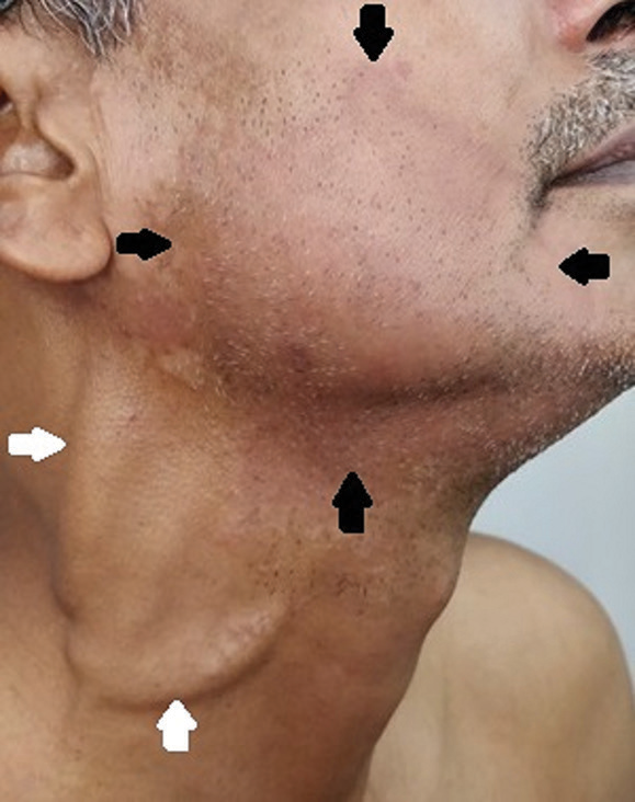
- Cord like, swellings on the right side of neck (white arrow), and hypoesthetic, erythematous patch on the right side of the lower cheek extending to the neck (borders marked by black arrows) in a patient with borderline tuberculoid leprosy.
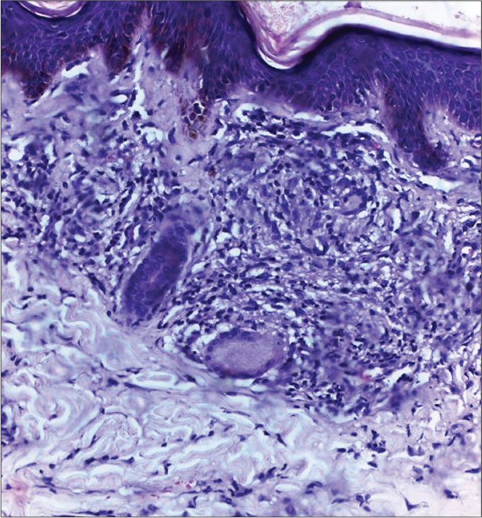
- Skin biopsy from the hypoesthetic, erythematous patch on the lower half of cheek showing epithelioid granuloma impinging on the epidermis, Langhan’s giant cells, and intra-granuloma and dermal edema suggestive of borderline tuberculoid leprosy with type 1 lepra reaction (H and E, ×200).
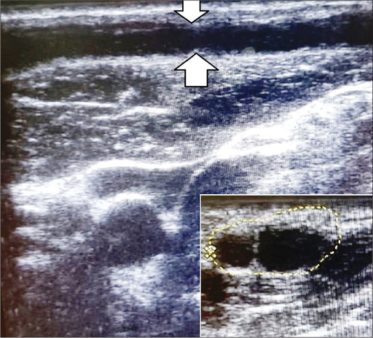
- High resolution ultrasound imaging done using 8–18 MHZ linear transducer of Esaote ultrasound machine in a patient with borderline tuberculoid leprosy with type 1 lepra reaction: Longitudinal sonogram showing linear hypoechoic structure suggestive of enlarged great auricular nerve (area between the white arrows); inset: Transverse sonogram showing two well defined, oval, hypoechoic structures suggestive of enlarged fascicles (encircled area) of great auricular nerve (cross-sectional area 55 mm2).
Ethical approval
The Institutional Review Board approval is not required.
Declaration of patient consent
The authors certify that they have obtained all appropriate patient consent.
Conflicts of interest
Dr. Sarita Sasidharanpillai is on the editorial board of the Journal.
Use of artificial intelligence (AI)-assisted technology for manuscript preparation
The authors confirm that there was no use of artificial intelligence (AI)-assisted technology for assisting in the writing or editing of the manuscript and no images were manipulated using AI.
Financial support and sponsorship
Nil.


