
Translate this page into:
Jonathan Hutchinson – A multispecialist of medicine
*Corresponding author: Vishalakshi S. Pandit, Department of Dermatology, Venereology and Leprosy, Koppal Institute of Medical Sciences, Koppal, Karnataka, India. vishalaxisp@gmail.com
-
Received: ,
Accepted: ,
How to cite this article: Pandit VS. Jonathan Hutchinson – A multispecialist of medicine. J Skin Sex Transm Dis 2024:6:22-5. doi: 10.25259/JSSTD_4_2024
Abstract
Sir Jonathan Hutchinson was a renowned dermatologist, surgeon, syphilologist, neurologist, and medical educator. Although his fields of interest were medicine, surgery, and ophthalmology, he contributed tremendously in the field of dermatology. He had made many original observations and published articles; many of the signs and diseases are named after him. A few to name are Hutchinson teeth, triad, nail sign, facies. Along with being a prolific author, he was a collector and teacher of many subjects. He had a feather on cap of publishing more than 600 articles and case reports in dermatology. He wrote the 10 volumes of Archives of Surgery, a remarkable singlehanded labor. He had earned honorary degrees from Universities of Glasgow, Cambridge, Edinburgh, Oxford, Dublin, and Leeds. He was knighted in 1908.
Keywords
Syphilis
Progeria
Hutchinson’s teeth

Sir Jonathan Hutchinson was a renowned dermatologist, surgeon, syphilologist, neurologist, and medical educator. He was born in 1828 in Selby, Yorkshire, the second of a family of 12 children. His field of interest was not only dermatology but also medical, surgical, neurological, and ophthalmic subjects. He made a tremendous contribution to the field of dermatology.[1] He was also a prolific writer of his time and contributed to “The Archives of Surgery” and the British Medical Journal. He made many original observations and published more than 600 articles and case reports in the field of dermatology. Many of the signs and diseases are named after him. A list of eponymous signs and diseases is shown in Table 1:
| S. No. | Disease/Condition |
|---|---|
| 1. | Hutchinson’s angina |
| 2. | Hutchinson–Boeck disease |
| 3. | Hutchinson’s facies |
| 4. | Hutchinson’s dyshidrosis |
| 5. | Hutchinson’s disease or senile degeneration of the choroid |
| 6. | Hutchinson–Gilford progeria syndrome |
| 7. | Hutchinson–Horton syndrome |
| 8. | Hutchinson’s melanotic disease |
| 9. | Hutchinson melanotic freckle |
| 10. | Hutchinson’s patch |
| 11. | Hutchinson’s prurigo |
| 12. | Hutchinson’s pupil |
| 13. | Hutchinson’s sign |
| 14. | Hutchinson’s teeth |
| 15. | Hutchinson’s triad |
Hutchinson’s angina:[2] Angioma serpiginosum is a rare vascular disorder with a characteristic appearance of clusters of tiny punctate telangiectasias in serpiginous pattern with a predilection for the extremities [Figure 1]. It usually affects females and starts during the first two decades of life.[3]
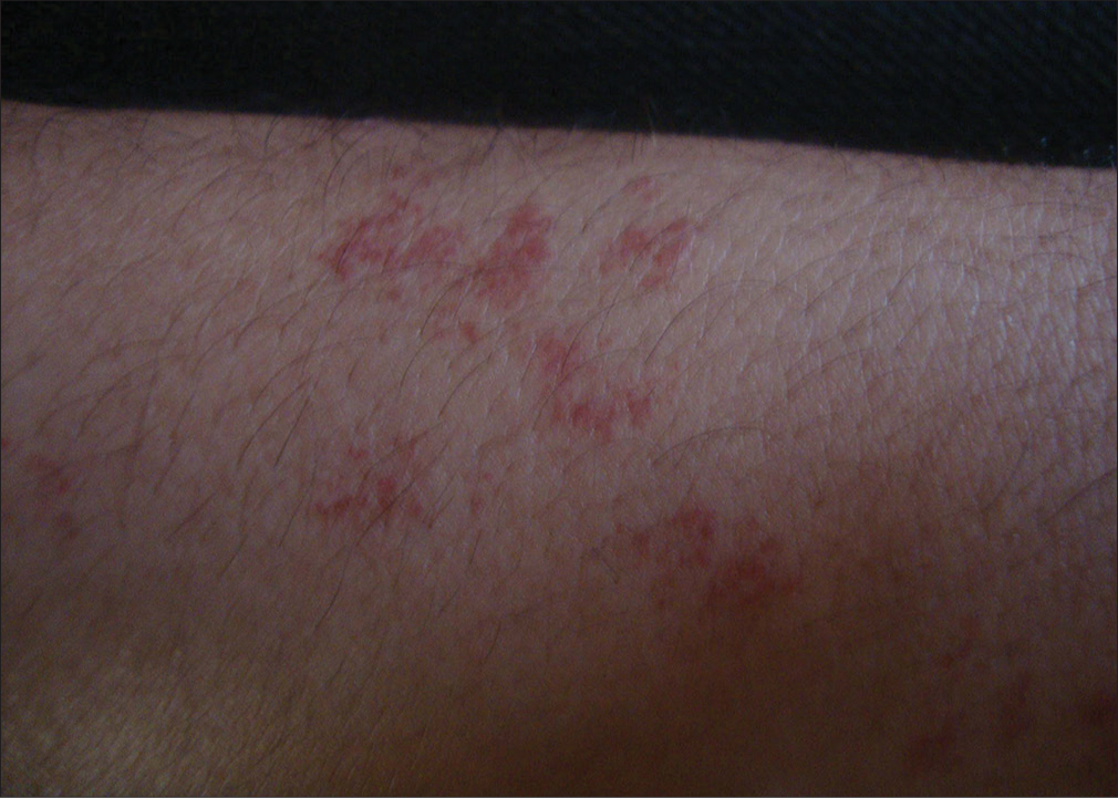
- Angioma serpiginosum: Multiple pinpoint coppery red maculopapular eruption on the side of a forearm
Hutchinson-Boeck disease: Earlier known as Mortimer’s malady (named after the patient’s name), credited to Hutchinson, who described the first case of sarcoidosis in his Illustrations of Clinical Surgery. He had described his case as “lupus vulgaris multiplex non-ulcerans” (Mortimer’s malady) to differentiate it from classical lupus vulgaris. FT Hunter and other authors later concluded that this was the first case of sarcoidosis.[4] His case had multiple, raised, dusky-red patches, which showed no tendency to inflame or ulcerate. The lesions appeared in groups and showed slow extension but were persistent and symmetrical in distribution.
Hutchinson’s facies: Mask-like face (flattening of facial lines) due to flabbiness of facial muscles and ptosis constitutes Hutchinson facies in tabes dorsalis.[5]
Hutchinson’s dyshidrosis: Cheiropompholyx was described by Hutchinson as the rapid and symmetrical development of vesicles [Figure 2], which tend to cure spontaneously and recur over again and again, mainly on the palms, later on, soles and sometimes, on other parts of the body. These eruptions usually start with intense burning and itching sensation of the hands.[6]
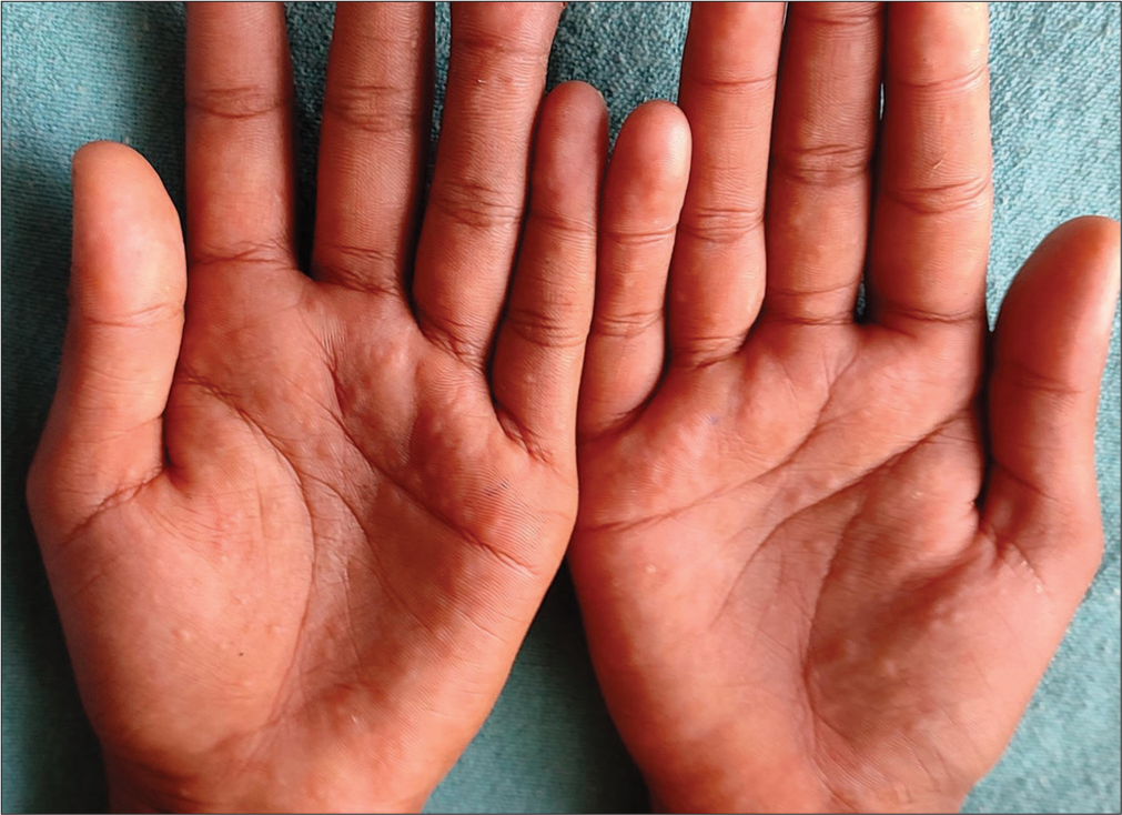
- Cheiropompholyx showing confluent vesicles on both palms.
Hutchinson–Gilford progeria syndrome: It is a rare premature aging disease that usually manifests at 6–18 months of life with cutaneous features such as perioral and nasolabial cyanosis, dyspigmentation, alopecia of scalp and eyebrows, early skin wrinkling, osteopenia, midfacial hypoplasia, cardiomegaly, angina, and myocardial infarction.[7]
Hutchinson-Horton syndrome: Hutchinson described a case of temporal arteritis presenting in an older adult as painful red streaks over the head; pulsations of these vessels were feeble, later ceasing to leave resistant cords. There was no history of headaches. Systemic manifestations of this condition include anorexia, insomnia, weight loss, low-grade fever, and sometimes, blindness may result if there is involvement of an ophthalmic artery.[8]
Hutchinson’s melanotic disease: It is also known as melanotic whitlow of Hutchinson. It is a rare melanotic tumor of the nail bed associated with melanotic tissue forming under and around the nail. It is of unknown etiology, presenting in middle to old age, and characterized by a pigmented band on the nail or chronic paronychia or warty growth on the nail bed.[9] It is also described as Hutchinson’s sign of nail where there is the presence of longitudinal melanonychia with the extension of hyperpigmentation onto the lateral and proximal nail fold representing the periungual spread of the in situ component of melanoma of nail matrix.[10]
Hutchinson’s melanotic freckle: Often known as the malignant freckle of Hutchinson. Hutchinson described the evolution of pigmented spots with malignant potential as follows: Usually appearing as brown spots on the eyelids of elderly people, later becoming black and enlarging in size. Then, multiple freckles coalesce and spread to adjacent mucous membranes such as the cornea and become prone to infections. In the later stage, growths may appear, which are non-pigmented, often break down to form ulcers and spread to adjacent tissues. Such cases were not limited just to eyelids but had been described on the lips and prepuce as well.[11] Now, this condition is recognized as lentigo maligna.
Hutchinson’s patch: Salmon-colored area in the cornea of syphilitic keratitis. The pathogenesis of syphilitic stromal keratitis is hypothesized to be the corneal autoimmune reaction involving antigenic mimicry.[12]
Hutchinson’s prurigo: It is also known as hydroa aestivale/actinic prurigo. It is a chronic familial photodermatosis of childhood, characterized by the presence of persistent pruritic, excoriated papular, and nodular lesions on the sun-exposed areas of the body. It is a persistent, distinct variant of polymorphic light eruption and is usually worse in summer.[13]
Hutchinson’s pupil: The fixed dilated pupil on the side of the temporal lobe coning occurs in the parenchymatous stage of neurosyphilis. Other causes for fixed dilated pupils are encephalitis, diabetes mellitus, multiple sclerosis, and trauma.[14]
Hutchinson’s sign: The involvement of the nasociliary branch of the ophthalmic division of the trigeminal nerve by varicella zoster virus presents with vesicular eruptions on the tip of the nose [Figure 3]. This indicates the involvement of intraocular structures of that side as the nasociliary branch innervates the eye and side and tip of the nose. Hence, Hutchinson stated that careful attention must be given to the eye if herpes zoster ophthalmicus is diagnosed with the involvement of the side and tip of the nose.[15] Involvement of the nasociliary branch is frequently accompanied by unilateral conjunctivitis and impaired corneal sensation, which can lead to corneal ulceration and sight-threatening bacterial infection.
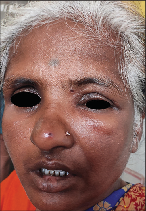
- Clinical photograph of herpes ophthalmicus showing vesicle on the tip of the nose.
Hutchinson’s teeth: The upper central incisors in congenital syphilis are characteristically narrow and short. The narrowness is at the free edge rather than at the crown, and a deep vertical notch is seen in the center of the free edge due to the breaking away or non-development of the middle lobe of the tooth crown. These rounded peg-shaped teeth may have a semi-translucent appearance.[1]
Hutchinson’s triad: Hutchinson established the importance of a triad of interstitial keratitis, congenital syphilitic teeth, and labyrinthine disease in the diagnosis of congenital syphilis.[11] He was also the first to declare that syphilis was due to a specific organism introduced into the body as a contagion.[1] He described the manifestations of late congenital syphilis, such as scars (rhagades) resulting from cutaneous fissures; a saddle-nose deformity resulting from destruction of nasal cartilage; anterior bowing of the mid tibia (saber shins); and scaphoid scapula, chronic large joint pain all resulting from chronic periostitis. In addition, he explained the varied skin manifestations of syphilis presenting in three stages and internal organ involvement and called it a great imitator and treated the disease with mercury pills and iodides.[1]
Hutchinson described lymphangioma circumscriptum as lupus lymphaticus [Figure 4]. His cases showed a warty appearance, initially developing as vesicles with surrounding thickened skin. Later, these vesicles became firm and very persistent, often discharged fluid, and had frequent episodes of erysipelatous inflammation. Hutchinson treated such cases with the application of caustics and actual cautery.[11]
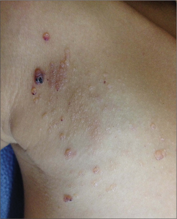
- Lymphangioma circumscriptum showing multiple vesicles on the right side of the chest.
Apart from these eponyms, he also contributed to many original descriptions of diseases in dermatology. A few of them are as follows:
Solid edema of the face following attacks of lymphangitis, a form of elephantiasis[11]
Atrophic balanitis[16]
Recurrent fibroid of the skin, a rare form of spindle cell sarcoma[11]
Penman’s disease, severe acne which involutes to form scars[11]
Severe bullous eruption produced by potassium iodide[11]
Varicella gangrenosa, also dermatitis gangrenosa infantum[16]
Arsenical keratoses and its oncogenic effects[16]
Crateriform ulcer on face (keratoacanthoma).
Ethical approval
Institutional Review Board approval is not required.
Declaration of patient consent
The authors certify that they have obtained all appropriate patient consents.
Conflicts of interest
There are no conflicts of interest.
Use of artificial intelligence (AI)-assisted technology for manuscript preparation
The authors confirm that there was no use of artificial intelligence (AI)-assisted technology for assisting in the writing or editing of the manuscript and no images were manipulated using AI.
Financial support and sponsorship
Nil.
References
- Eminent venereologists 4: Jonanthan Hutchinson. Genitourin Med. 1990;66:401-6.
- [CrossRef] [PubMed] [Google Scholar]
- Eponyms of Sir Jonathan Hutchinson. Int J Dermatol. 2008;47:754-8.
- [CrossRef] [PubMed] [Google Scholar]
- Diseases of the veins and arteries: Leg ulcers In: Burns T, Breathnach S, Cox N, Griffiths C, eds. Rook's textbook of dermatology (8th ed). United States: Wiley-Blackwell; 2010. p. :471-58.
- [CrossRef] [PubMed] [Google Scholar]
- Clinical manifestations of syphilis In: Holmes KK, Sparling PF, Stamm WE, Piot P, Wasserheit JN, Corey L, eds. Sexually transmitted diseases (4th ed). New York: McGraw-Hill Companies; 2008. p. :661-84.
- [Google Scholar]
- Phenotype and course of Hutchinson-Gilford progeria syndrome. N Engl J Med. 2008;358:592-604.
- [CrossRef] [PubMed] [Google Scholar]
- The history of Horton's disease or. 10 centuries of a fascinating adventure. J Mal Vasc. 1989;14(Suppl C):93-7.
- [Google Scholar]
- Melanotic disease of the great toe following a whitlow of the nail. Trans Pathol Soc Lond. 1857;8:404-5.
- [Google Scholar]
- Nail disorders In: Kang S, Amagai M, Bruckner AL, Enk AH, Margolis DJ, McMichael AJ, eds. Fitzpatrick's dermatology (9th ed). United States: McGraw-Hill Education; 2019. p. :1606-8.
- [Google Scholar]
- Dermatological writings of Sir Jonathan Hutchinson. AMA Arch Derm Syphilol. 1952;65:130-6.
- [CrossRef] [PubMed] [Google Scholar]
- Adult-onset syphilitic stromal keratitis. Am J Ophthalmol. 2006;141:319-21.
- [CrossRef] [PubMed] [Google Scholar]
- Photodermatology and photodermatoses In: Sacchidanand S, Savitha AS, Shilpa K, Shashikumar BM, eds. IADVL textbook of dermatology (5th ed). Mumbai: Bhalani Publishers; 2021. p. :887-929.
- [Google Scholar]
- The differential diagnosis of fixed dilated pupils: A case report and review. Crit Care Resusc. 2000;2:34-7.
- [CrossRef] [PubMed] [Google Scholar]
- Human herpes viruses In: Bolognia JL, Schaffer JV, Cerroni L, eds. Dermatology (4th ed). China: Elsevier; 2018. p. :1400-22.
- [Google Scholar]
- Hutchison's archives of surgery revisited. Arch Dermatol. 1997;113:961-4.
- [CrossRef] [Google Scholar]


