Translate this page into:
Lupus in the guise of pure neuritic leprosy
*Corresponding author: Raman Balakrishna Venkatta Department of Health Services, Palakkad, Kerala, India. doctor82@gmail.com
-
Received: ,
Accepted: ,
How to cite this article: George AE, Venkatta RB. Lupus in the guise of pure neuritic leprosy. J Skin Sex Transm Dis 2020;2(2):122-5.
Abstract
Nerve palsy without skin lesions would bring to mind, pure neuritic form of leprosy in endemic countries like India. However, it is imperative to keep in mind the possibilities of other conditions like systemic lupus erythematosus; the misdiagnosis of which can lead to disastrous consequences. Here, we discuss such a case which was initially suspected to be pure neuritic leprosy and later diagnosed just on time for emergency management of eye complications.
Keywords
Leprosy
systemic lupus erythematosus
Neuritis
INTRODUCTION
Peripheral nerve involvement with nerve thickening and/or skin lesions is commonly seen in leprosy and less commonly in other conditions. Systemic lupus erythematosus (SLE) is a multisystem disorder which may involve the central nervous system (CNS) also. The involvement of peripheral nerves in SLE is seen in 2–18%.[1] The onset of nerve involvement is insidious in conditions like leprosy while it may be acute and often pose a diagnostic dilemma in SLE. Awareness and recognition of SLE as a cause of acute peripheral nerve palsy warrants emergency intervention to avoid permanent damage, especially as in the case of optic nerve involvement. A case of a young female, initially misdiagnosed as pure neuritic leprosy (PNL), is presented here.
CASE REPORT
A 17-year-old girl was brought to the outpatient wing of the department of dermatology and venereology with complaints of weakness of both hands for the past 2 weeks. She was referred by a dermatologist with request to do a nerve biopsy to confirm a diagnosis of PNL with Type 2 lepra reaction. She had a history of intermittent fever with intermittent episodes of abdominal pain and multiple joint pain for the past 1 month. As the patient was found to have positive serological tests for enteric fever, she had been treated with ceftriaxone from the referring hospital. There was no history of contact with patients having leprosy.
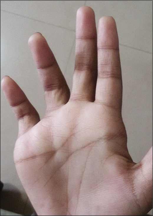
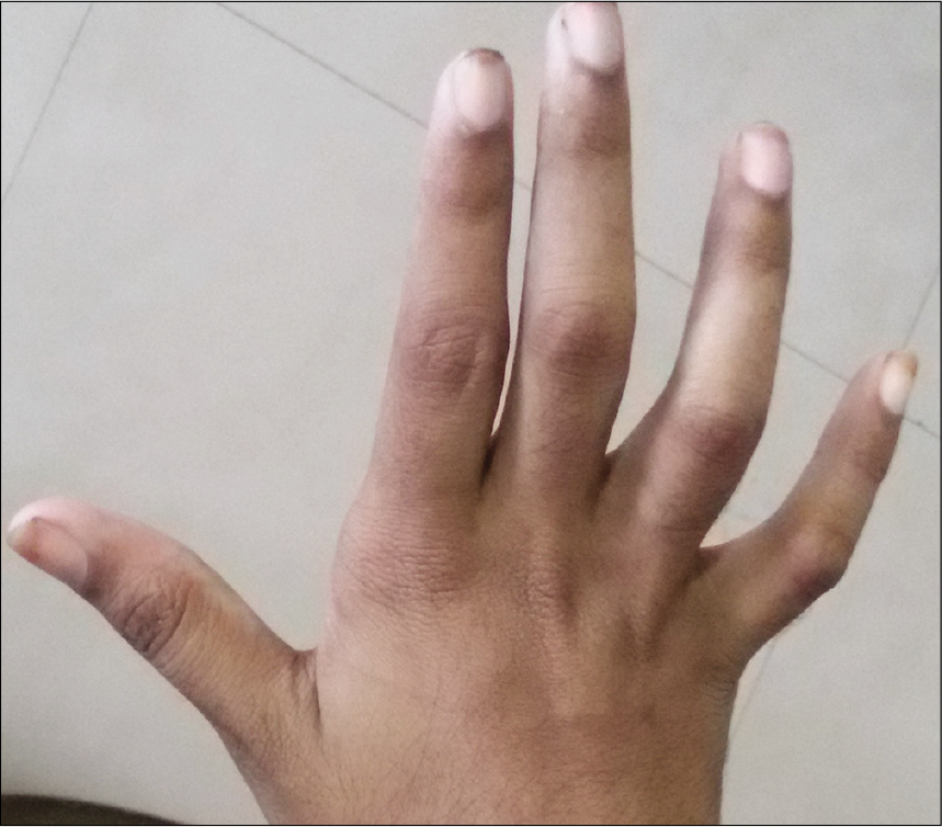
On examination, she was febrile and pale. There was ulnar clawing of the right hand [Figures 1 and 2] and her right ulnar nerve was slightly thickened, non-tender, and non-nodular. The right radial cutaneous nerve was slightly tender without any nodularity. Temperature, touch, and pain perceptions were impaired over her right hand in an ulnar distribution. Motor system examination revealed positive book test, card test, and negative pen test. She had post-inflammatory hyperpigmentation following varicella zoster 18 months back and her dermatological examination was otherwise unremarkable. On routine blood examination, she had anemia (Hb – 7.8 mg/dL), erythrocyte sedimentation rate – 21 mm/h, and hypoalbuminemia (2.6 mg/dL). ELS/SSS was negative for Mycobacterium leprae. Anti-nuclear antibody (ANA) report was pending.

- Claw hand.

- Dorsal view of claw hand.
With a provisional diagnosis of PNL, she was admitted for further investigations including nerve biopsy and histopathology analysis. A possibility of Type 1 lepra reaction was also considered in view of the history of intermittent fever for the past 1 month and tenderness along the course of a peripheral nerve, i.e., the right radial cutaneous nerve. However, the ulnar nerve was non-tender.
On the night of the admission, the patient complained of sudden, painless loss of vision of her right eye. Examination revealed a relative afferent pupillary defect and emergency ophthalmology and neurology consultations were done. In view of the intermittent fever, joint pain, recent acute onset of multiple nerve involvement, and investigation results, the possibility of SLE was considered and she was immediately started on I.V. methyl prednisolone in an attempt to save her eyesight.
Her ANA result turned out to be positive. Further investigations revealed positive anti-dsDNA, Scl-70, PMScl, anti-histones, and AMA-M2. Peripheral smear showed microcytic hypochromic anemia. Other hematological parameters were within normal limits. USG revealed hepatomegaly with minimal free fluid in the pouch of Douglas.
Visual evoked potential recording showed prolonged P100 latency in the left eye and was normal in the right eye. The amplitude in both eyes was reduced with an interocular asymmetry R>L.
Nerve conduction study of upper limb showed that the right ulnar F-wave was absent and there was reduced amplitude in the left median nerve (1.3 mv). Sensory conduction showed absent sensory nerve action potentials (SNAPs) from the right ulnar nerve and dorsal ulnar cutaneous nerve. Compound muscle action potential was not elicitable from the right ulnar nerve at all levels. There was asymmetry between bilateral SNAP amplitudes of median nerves (L – 13.8 μv vs. R – 34 μv). Slit skin smear for acid-fast bacilli (AFB) taken from anesthetic area and normal skin, ear lobe smear for AFB, and histopathology from the anesthetic area were unremarkable. The patient did not give consent for nerve biopsy in view of potential to cause further damage to the motor nerve.
The patient responded favorably to the treatment and regained eyesight the very next day. It was only 2 days after starting IV prednisolone that she developed faint erythematous rash over the malar area. Skin biopsy was not taken from the face as the diagnosis was already established with serology and systemic features. However, there was hardly any improvement to the claw hands.
DISCUSSION
The diagnosis of PNL remains a public health care problem in endemic countries like India because skin lesions are never present. Investigations like FNAC from the involved nerve and even a biopsy from the nerve may not result in identification of the leprosy bacillus. In developing countries, leprosy is the most common cause of peripheral neuropathy and a provisional diagnosis of PNL was well justified in this case.[2]
SLE is a chronic, idiopathic, and multisystem inflammatory disease characterized by autoantibody production and hyperactivity of the immune system. The diagnosis can be confirmed if 4 of 11 American College of Rheumatology (ACR) criteria are met. SLE may affect the eyes and/or visual system in up to a third of patients and ocular involvement in SLE may be the presenting feature of the disease and is still a potentially blinding condition.[3]
The peripheral neuropathy in SLE may be of progressive symmetric sensorimotor type, motor neuropathy, multiple mononeuropathy, or a radiculoneuropathy. The ACR nomenclature for neuropsychiatric SLE provides case definitions for neuropsychiatric syndromes seen in SLE.[4] The most common clinical presentation of peripheral neuropathy was a distal axonal sensory or sensory-motor polyneuropathy with acute or subacute onset. An asymmetrical involvement of the extremities was the most frequent presentation in many studies.[5]
The cranial nerves, being peripheral nerves, may be affected in leprosy, but the optic nerve does not appear to be affected in patients with leprosy.[6] Even amidst the plethora of ocular complications of leprosy, optic nerve involvement in leprosy is considered extremely rare.[7] Sudden painless vision loss can occur in other conditions such as amaurosis fugax, giant cell arteritis, central retinal artery occlusion, central retinal vein occlusion, vitreous hemorrhage, ischemic optic neuropathies, posterior cerebrovascular accidents, and retinal detachment.[8]
Ocular lesions are not included among the diagnostic criteria of SLE and many researchers believe that it is an oversight. Inclusion of ocular disease would lead to earlier diagnosis and therapeutic intervention in those instances.[9] Optic nerve involvement, however, occurs in only up to 1% of patients with SLE. The clinical picture is variable and may be insidious with slowly progressive visual loss or can present as acute retrobulbar optic neuritis, ischemic optic neuropathy, afferent pupillary nerve defect, visual field loss or scotoma, and normal-appearing or swollen optic disc.[10] Despite the variable presentations, the probable pathogenesis in all cases is vaso-occlusive disease in small vessels of the optic nerves, by immune complex with antiphospholipid deposition, vasculitis, thrombosis, or infections.[11] Fundus changes in SLE may confuse the physician with that of concomitant hypertension but can represent an independent effect of the disease process.[12] Retinopathy is a marker of disease activity and CNS involvement. It can be a guide in the management of patient, but other causes of the ocular illness have to be ruled out.[13]
Peripheral neuropathy improved in 65.8% of patients with SLE in a study.[5] The prognosis in individuals with SLE and optic neuropathy is poor because it is notoriously difficult to treat. The visual outcome varies, but improvement occasionally occurs following treatment with corticosteroids. The standard treatment is corticosteroid therapy either orally or pulsed. However, recovery, if any, is slow with final visual acuity of 20/200 or worse in about 55% of patients as reported in a series.[10]
Although our patient did not have any skin lesions suggestive of Hansen’s disease, the occurrence of motor and sensory weakness and claw hand in the patient who had been living in an area endemic for leprosy raised the possibility of pure neuritic Hansen’s disease at first. Moreover, the history of intermittent fever, nerve tenderness, and arthralgia was suggestive of Type 1 lepra reaction. However, the acute onset of optic nerve involvement with sudden loss of vision raised the suspicion of other possible diagnoses like SLE which was confirmed on investigation and prompt treatment with systemic corticosteroids averted a major disaster like permanent loss of vision. The patient had all features to diagnose lupus. She had only slight thickening of her right ulnar nerve which was non-tender and non-nodular. Sudden onset nerve palsy as observed in this case is seen in lepra reaction usually, but such sudden onset nerve function impairment is associated with nerve tenderness. In this case, the right ulnar nerve which developed sudden onset nerve palsy had no tenderness and, therefore, was not suggestive of Type 1 reaction. A marginally thickened, but non tender, right ulnar nerve could be a normal finding in a person with right hand dominance. That could explain the thickened ulnar nerve in our patient and it was unlikely to be due to leprosy. The electrophysiological studies of the ulnar nerve were also of neuropathy seen in lupus, but it cannot be differentiated from that in leprosy. She had also not developed any other nerve involvement or skin lesions suggestive of leprosy during her follow-up in the next 8 months.
CONCLUSION
Although the presence of isolated nerve deficit in patients from leprosy endemic areas should raise the suspicion of PNL, it is always important to keep all the other differential diagnoses in mind, especially when unusual symptoms arise.
Declaration of patient consent
The authors certify that they have obtained all appropriate patient consent.
Financial support and sponsorship
Nil.
Conflicts of interest
Dr. Anuja Elizabeth George is on the Editorial Board of the Journal.
References
- Peripheral sensorimotor and autonomic neuropathy associated with systemic lupus erythematosus: Clinical, pathological and immunological features. Brain. 1987;110:533-49.
- [CrossRef] [PubMed] [Google Scholar]
- Criteria for diagnosis of pure neural leprosy. J Neurol. 2003;250:806-9.
- [CrossRef] [PubMed] [Google Scholar]
- Ocular manifestations of systemic lupus erythematosus. Rheumatology. 2007;46:1757-62.
- [CrossRef] [PubMed] [Google Scholar]
- The American College of Rheumatology nomenclature and case definitions for neuropsychiatric lupus syndromes. Arthritis Rheum. 1999;42:599-608.
- [CrossRef] [Google Scholar]
- Peripheral neuropathy in patients with systemic lupus erythematosus. Semin Arthritis Rheum. 2011;41:203-11.
- [CrossRef] [PubMed] [Google Scholar]
- Bacteria and bacterial diseases In: Miller NR, Nancy J, eds. Walsh and Hoyt's Clinical Neuro-ophthalmology (6th ed). United States: Lippincott Williams & Wilkins; 2005. p. :2647-763.
- [Google Scholar]
- Optic nerve involvement in a borderline lepromatous leprosy patient on multidrug therapy. Lepr Rev. 2013;84:316-21.
- [Google Scholar]
- Ocular manifestations of systemic lupus erythematosus. Curr Opin Ophthalmol. 2002;13:404-10.
- [CrossRef] [PubMed] [Google Scholar]
- Clinical aspects of the nervous system. In: Dubois Lupus Erythematosus Related Syndromes, Expert Consult-Online. Vol 24. Australia: Saunders; 2012. p. :368.
- [CrossRef] [Google Scholar]
- Optic neuropathy in systemic lupus erythematosus. Arch Ophthalmol. 1986;104:564-8.
- [CrossRef] [PubMed] [Google Scholar]
- Ocular findings in systemic lupus erythematosus. Br J Ophthalmol. 1972;56:800-4.
- [CrossRef] [PubMed] [Google Scholar]
- Lupus retinopathy, Patterns, associations, and prognosis. Arthritis Rheum. 1988;31:1105-10.
- [CrossRef] [PubMed] [Google Scholar]


